. 2014 Sep 15:4:6356.
doi: 10.1038/srep06356.

Characterizing the pocketome of Mycobacterium tuberculosis and application in rationalizing polypharmacological target selection

Affiliations

Characterizing the pocketome of Mycobacterium tuberculosis and application in rationalizing polypharmacological target selection

Praveen Anand et al. Sci Rep. .

Abstract

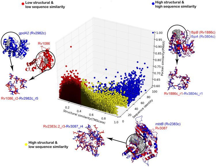
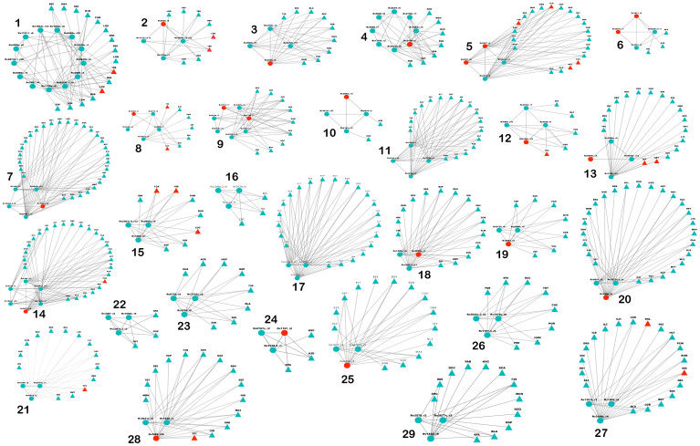
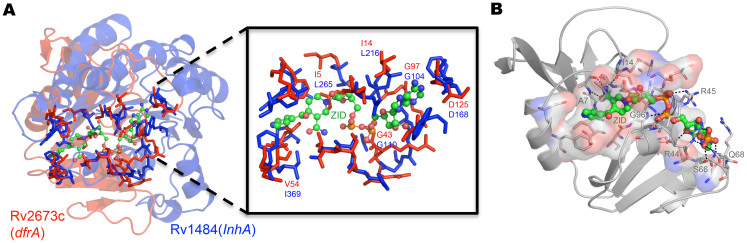
Polypharmacology is beginning to emerge as an important concept in the field of drug discovery. However, there are no established approaches to either select appropriate target sets or design polypharmacological drugs. Here, we propose a structural-proteomics approach that utilizes the structural information of the binding sites at a genome-scale obtained through in-house algorithms to characterize the pocketome, yielding a list of ligands that can participate in various biochemical events in the mycobacterial cell. The pocket-type space is seen to be much larger than the sequence or fold-space, suggesting that variations at the site-level contribute significantly to functional repertoire of the organism. All-pair comparisons of binding sites within Mycobacterium tuberculosis (Mtb), pocket-similarity network construction and clustering result in identification of binding-site sets, each containing a group of similar binding sites, theoretically having a potential to interact with a common set of compounds. A polypharmacology index is formulated to rank targets by incorporating a measure of druggability and similarity to other pockets within the proteome. This study presents a rational approach to identify targets with polypharmacological potential along with possible drugs for repurposing, while simultaneously, obtaining clues on lead compounds for use in new drug-discovery pipelines.


Conflict of interest statement

The authors declare no competing financial interests.

Figures

Figure 1
Figure 1. An overview of the characterization of the enzymes in the Mtb pocketome, in terms of binding site analysis.
(A) A stacked bar plot showing the coverage of protein structures and the confident ligand associations available with respect to the KEGG pathways. For each pathway the lower most bar in the stack corresponds to the number of genes or proteins in the pathway, the middle bar indicates the number of structural models available for the pathway and the top most stack indicates the number of proteins for which ligand annotations are made based on binding site structures. Each stack corresponds to one KEGG pathway in Mtb. (B) Metabolic map of central metabolism in Mtb, indicating extensive coverage of ligand annotation in the Mtb reactome from this study. The edges colored in black indicates the availability of protein structure catalyzing the reaction and the nodes colored in red represent the small ligand molecules taking part in the reaction for which the binding site has been mapped onto the respective protein structure.
Figure 2
Figure 2. An illustration of ligand associations for Mtb pocketome.
Distribution of different ligand hits obtained for the predicted pockets in the proteome. The ligands are ordered by their molecular weights. The frequency on the Y-axis indicates the number of occurrences of the binding site of that ligand in the Mtb pocketome. This spectrum is qualitatively equivalent to the mass spectrum of the Mtb metabolome for unit protein abundances.
Figure 3
Figure 3. Binding Site Similarity networks.
(A) The binding site similarity network obtained for Mtb Pocketome. Each node represents the predicted binding site and an edge between two nodes represents high similarity shared (PMAX ≥ 0.7) between them. The colors represent different clusters or sets of binding sites predicted by MCODE algorithm. (B) Binding site similarity network of pockets obtained from MOAD dataset, carried out as a validation exercise. The color of the nodes again depicts set of similar binding sites obtained from MCODE algorithm. Three such example clusters binding to ATP, heme and phosphoglycerate respectively are shown in enlarged version.
Figure 4
Figure 4
An overview of all-pair binding site similarities in Mtb Pocketome representing the results of 96 million comparisons (A) Hexbin plot depicts the distribution of all-pair similarity scores obtained using PocketMatch. The y-axis depicts the local or partial binding site similarity scores (PMIN) and the x-axis depicts the global-similarity scores (PMAX). The color of the hexbin represents the density of the scores obtained and is shown in the legend next to the plot. (B) Distribution of all-pair PMIN scores. (C) Distribution of all-pair PMAX scores. (D) Degree distribution of the sites in Mtb binding site similarity network, indirectly capturing number of similar sites.
Figure 5
Figure 5. An illustration of the structure-sequence-pocket space relationships in Mtb proteome.
The 3D scatterplot depicts the distribution of high similarity pockets with respect to sequence and structural similarity scores obtained for the corresponding proteins. The color represents different categories of sequence-structure relationship and an example is highlighted from each of these categories with the depiction of proteins and pockets similarity.
Figure 6
Figure 6. Drug-hits for Polypharmacological targets.
Each disconnected component represents a set of polypharmacological targets obtained from Mtb binding site similarity network. Two type of nodes are present in the network, the predicted binding sites are shown as spheres and the drugs sharing a binding site similarity are shown as triangles. The red colored circular nodes represent binding sites of high-confidence targets. Approved drugs are also highlighted in red.
Figure 7
Figure 7
A selected example of similar binding sites in different proteins predicted in this study matching with crystal structures available in literature (A) Structural superposition of dihydrofolate reductase (red cartoon) and InhA (blue cartoon, PDBID: 1ZID) protein based on the similarity of the binding sites (shown as sticks). The inset shows the similarity of the binding sites with the isoniazid adduct shown in ball and stick representation. (B) Crystal structure of dihydrofolate reductase with characterization of binding site for isoniazid adduct (PDBID: 2CIG).

References

    1. Konopa K. & Jassem J. The role of pemetrexed combined with targeted agents for non-small cell lung cancer. Curr Drug Targets 11, 2–11 (2010). - PubMed
    1. Winum J. Y., Maresca A., Carta F., Scozzafava A. & Supuran C. T. Polypharmacology of sulfonamides: pazopanib, a multitargeted receptor tyrosine kinase inhibitor in clinical use, potently inhibits several mammalian carbonic anhydrases. Chem Commun (Camb) 48, 8177–9 (2012). - PubMed
    1. Hopkins A. L. Network pharmacology: the next paradigm in drug discovery. Nat Chem Biol 4, 682–90 (2008). - PubMed
    1. Csermely P., Korcsmaros T., Kiss H. J., London G. & Nussinov R. Structure and dynamics of molecular networks: a novel paradigm of drug discovery: a comprehensive review. Pharmacol Ther 138, 333–408 (2013). - PMC - PubMed
    1. Zhao S. et al. Systems pharmacology of adverse event mitigation by drug combinations. Sci Transl Med 5, 206ra140 (2013). - PMC - PubMed

Publication types

LinkOut - more resources