. 2014 Aug 4:20:1146-59.
eCollection 2014.

Role of FAM18B in diabetic retinopathy

Affiliations

Role of FAM18B in diabetic retinopathy

Ai Ling Wang et al. Mol Vis. .

Abstract

Purpose: Genome-wide association studies have suggested an association between a previously uncharacterized gene, FAM18B, and diabetic retinopathy. This study explores the role of FAM18B in diabetic retinopathy. An improved understanding of FAM18B could yield important insights into the pathogenesis of this sight-threatening complication of diabetes mellitus.

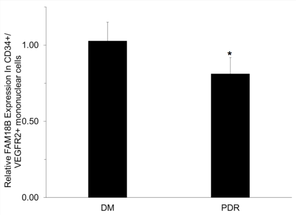
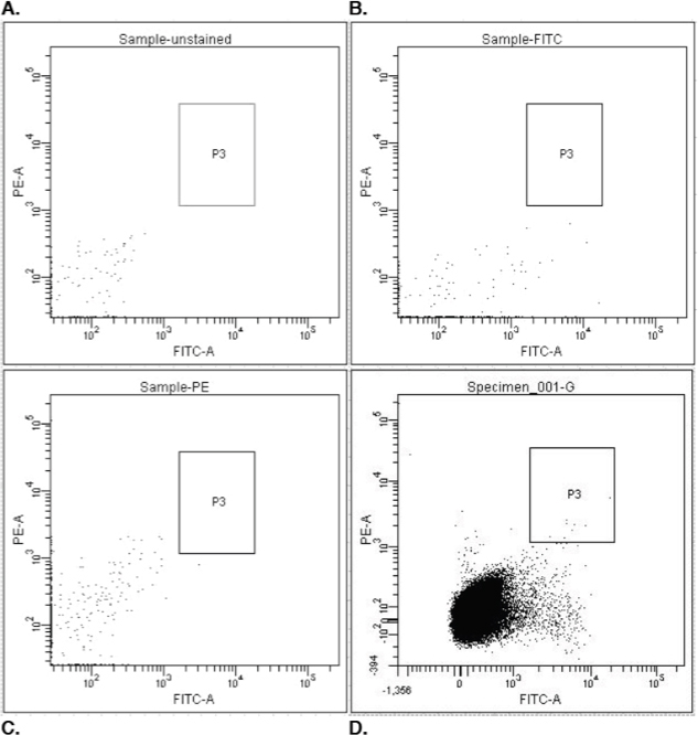
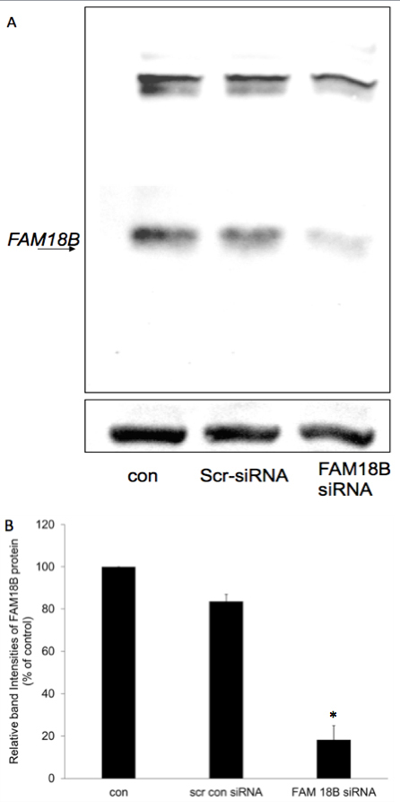
Methods: Postmortem human eyes were examined with immunohistochemistry and immunofluorescence for the presence of FAM18B. Expression of FAM18B in primary human retinal microvascular endothelial cells (HRMECs) exposed to hyperglycemia, vascular endothelial growth factor (VEGF), or advanced glycation end products (AGEs) was determined with quantitative reverse-transcription PCR (qRT-PCR) and/or western blot. The role of FAM18B in regulating human retinal microvascular endothelial cell viability, migration, and endothelial tube formation was determined following RNAi-mediated knockdown of FAM18B. The presence of FAM18B was determined with qRT-PCR in CD34+/VEGFR2+ mononuclear cells isolated from a cohort of 17 diabetic subjects with and without diabetic retinopathy.

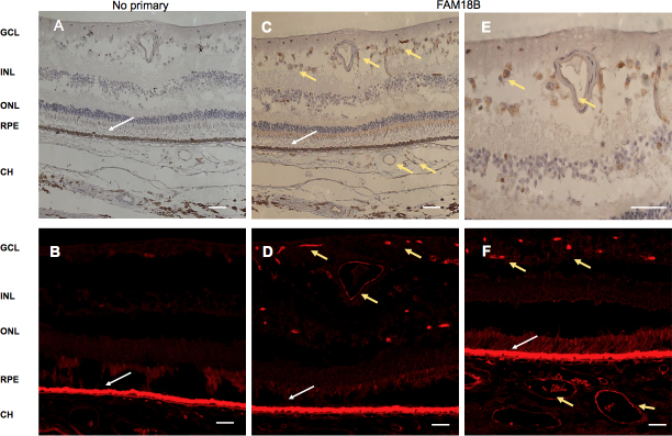
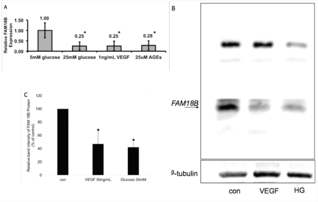
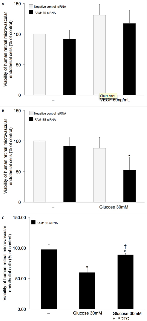
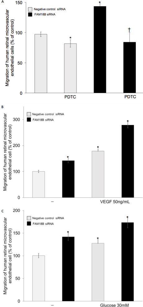
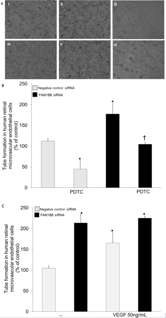
Results: Immunohistochemistry and immunofluorescence demonstrated the presence of FAM18B in the human retina with prominent vascular staining. Hyperglycemia, VEGF, and AGEs downregulated the expression of FAM18B in HRMECs. RNAi-mediated knockdown of FAM18B in HRMECs contributed to enhanced migration and tube formation as well as exacerbating the hyperglycemia-induced decrease in HRMEC viability. The enhanced migration, tube formation, and decrease in the viability of HRMECs as a result of FAM18B downregulation was reversed with pyrrolidine dithiocarbamate (PDTC), a specific nuclear factor-kappa B (NF-κB) inhibitor. CD34+/VEGFR2+ mononuclear cells from subjects with proliferative diabetic retinopathy demonstrated significantly reduced mRNA expression of FAM18B compared to diabetic subjects without retinopathy.

Conclusions: FAM18B is expressed in the retina. Diabetic culture conditions decrease the expression of FAM18B in HRMECs. The downregulation of FAM18B by siRNA in HRMECs results in enhanced migration and tube formation, but also exacerbates the hyperglycemia-induced decrease in HRMEC viability. The pathogenic changes observed in HRMECs as a result of FAM18B downregulation were reversed with PDTC, a specific NF-κB inhibitor. This study is the first to demonstrate a potential role for FAM18B in the pathogenesis of diabetic retinopathy.


Figures

Figure 1
Figure 1
Immunolocalization of FAM18B in the human retina. A, B: Negative controls for immunohistochemistry and immunofluorescence, respectively. Autofluorescence is evident in the RPE (white arrow). Immunohistochemistry demonstrates FAM18B (brown) in the retinal and choroidal vasculature (yellow arrows; C, E). Immunofluorescence reveals a prominent signal for the FAM18B (red) in the retinal and choroidal vasculature (yellow arrows; D, F).
Figure 2
Figure 2
Decreased FAM18B expression under diabetic conditions in cultured human retinal microvascular endothelial cells. A: There are significant decreases in the FAM18B mRNA levels in human retinal microvascular endothelial cells (HRMECs) treated with 25 mM glucose, 1 ng/ml vascular endothelial growth factor (VEGF), and 25 μM advanced glycation end products (AGEs), compared with 5 mM glucose. B: Western blot of the FAM18B protein and β-tubulin in HRMECs following treatment with VEGF 50 ng/ml and glucose 30 mM for 48 h reveals decreased protein expression. C: Quantification of the FAM18B protein band intensities normalized to β-tubulin band intensities in HRMECs following treatment with VEGF 50 ng/ml and glucose 30 mM for 48 h. Data are represented as percent change mean±standard deviation (SD) regarding control cells treated with media alone (n=3). * p value <0.05 compared to control.
Figure 3
Figure 3
Effect of FAM18B knockdown on human retinal microvascular endothelial cell viability. The viability of human retinal microvascular endothelial cells (HRMECs) in various treatment groups are expressed as the percent viability of HRMECs transfected with scrambled control siRNA and treated with medium alone unless specified otherwise. A: Viability of HRMECs transfected with scrambled control siRNA or FAM18B siRNA treated with or without vascular endothelial growth factor (VEGF). B: Viability of HRMECs transfected with scrambled control siRNA or FAM18B siRNA treated with or without high glucose. C: Viability of HRMECs transfected with FAM18B siRNA treated with high glucose in the presence or absence pyrrolidine dithiocarbamate (PDTC). AC: PDTC is a specific nuclear factor-kappa B (NF-κB) inhibitor. Data represented as mean±standard deviation (SD) (n=3×96-well plates; four wells/plate for panels B and C and n=1×96 well plate; five wells/plate for panel A). * p value <0.05 compared to control cells treated with media alone. †p value <0.05 compared to FAM18B siRNA-transfected cells treated with high glucose.
Figure 4
Figure 4
Effect of FAM18B knockdown on human retinal microvascular endothelial cell migration. The migration of human retinal microvascular endothelial cells (HRMECs) in various treatment groups are expressed as percent change of HRMECs transfected with scrambled control siRNA and treated with medium alone. A: Migration of HRMECs transfected with scrambled control siRNA or FAM18B siRNA in the presence or absence of pyrrolidine dithiocarbamate (PDTC). FAM18B knockdown significantly increases the basal migration of HRMECs. B: Migration of HRMECs transfected with scrambled control siRNA or FAM18B siRNA treated with or without vascular endothelial growth factor (VEGF). C: Migration of HRMECs transfected with scrambled control siRNA or FAM18B siRNA treated with or without high glucose. PDTC is a specific nuclear factor-kappa B (NF-κB) inhibitor. VEGF and glucose significantly increase the migration of control cells. The VEGF and glucose-induced migration is further potentiated in FAM18B siRNA-transfected cells. The data are represented as mean±standard deviation (SD) * p-value <0.05 compared to control cells treated with media alone. † p value <0.05 compared to FAM18B siRNA-transfected cells.
Figure 5
Figure 5
Effect of FAM18B knockdown on human retinal microvascular endothelial cell tube formation. A: Representative images of tube formation in various treatment groups. (i) Scrambled control; (ii) scrambled control treated with vascular endothelial growth factor (VEGF); and (iii) scrambled control treated with pyrrolidine dithiocarbamate (PDTC). (iv) FAM18B siRNA-treated cells; (v) FAM18B siRNA treated with VEGF; and (vi) FAM18B siRNA treated with PDTC. B: The tube formation of human retinal microvascular endothelial cells (HRMECs) in transfected with scrambled control siRNA or FAM18B transfected siRNA treated with or without PDTC. FAM18B knockdown increases the basal tube formation of HRMECs. The enhanced basal tube formation is inhibited by PDTC, a specific nuclear factor-kappa B (NF-κB) inhibitor. C: Tube formation in control siRNA-transfected cells and FAM18B siRNA-transfected cells treated with or without VEGF. VEGF increases tube formation of control cells but not in FAM18B siRNA transfected cells. The data are represented as mean±standard deviation (SD) (n=3). * p value <0.05 compared to control cells treated with media alone. † p value <0.05 compared to FAM18B siRNA-transfected cells.
Figure 6
Figure 6
Decreased FAM18B expression in CD34+/VEGFR2+ cells. Expression of FAM18B in CD34+/VEGFR2+ mononuclear cells from diabetic subjects with proliferative diabetic retinopathy is decreased compared to diabetic subjects without retinopathy. Glyceraldehyde 3-phosphate dehydrogenase (GAPDH) mRNA was used as internal standard for normalization. DM: diabetes without diabetic retinopathy (n=9). PDR: diabetes with proliferative diabetic retinopathy (n=8). *p-value <0.05.
Figure 7
Figure 7
Isolation of CD34+/VEGFR2+ mononuclear cells. Single positive controls and an unstained control were used to set the appropriate compensation values necessary to assign the double positive gating strategy (A, B, and C). Whole cells were separated from debris and doublets using the area and width pulses for forward and side scatter (D). The box identifies all double-positive cells that exhibit sufficient fluorescence of both markers to be identified—these were the cells that were sorted and collected.
Figure 8
Figure 8
RNAi-mediated downregulation of FAM18B in human retinal microvascular endothelial cells. A: Representative western blot of the FAM18B protein and β-tubulin in human retinal microvascular endothelial cells (HRMECs) following transfection with control siRNA and FAM18B siRNA for 72 h. B: Quantification of the FAM18B protein band intensities normalized to β-tubulin band intensities in HRMECs. Data are represented as percent change. Mean±standard deviation (SD) regarding cells treated with media alone (n=2). * p value <0.05.

References

    1. National diabetes fact sheet: national estimates and general information on diabetes and prediabetes in the United States. 2011 [cited 2012 02/02/2012]; Available from: http://www.cdc.gov/diabetes/pubs/pdf/ndfs_2011.pdf
    1. Tarr JM, Kaul K, Chopra M, Kohner EM, Chibber R. Pathophysiology of Diabetic Retinopathy. ISRN. 2013;2013:343560. - PMC - PubMed
    1. Klein R. Hyperglycemia and microvascular and macrovascular disease in diabetes. Diabetes Care. 1995;18:258–68. - PubMed
    1. Engerman RL, Kern TS. Hyperglycemia as a cause of diabetic retinopathy. Metabolism. 1986;35(Suppl 1):20–3. - PubMed
    1. Caldwell RB, Bartoli M, Behzadian MA, El-Remessy AE, Al-Shabrawey M, Platt DH, Caldwell RW. Vascular endothelial growth factor and diabetic retinopathy: pathophysiological mechanisms and treatment perspectives. Diabetes Metab Res Rev. 2003;19:442–55. - PubMed

Publication types

MeSH terms