

Lipid Droplets as Signaling Platforms Linking Metabolic and Cellular Functions
- PMID: 25221429
- PMCID: PMC4161058
- DOI: 10.4137/LPI.S11128
Lipid Droplets as Signaling Platforms Linking Metabolic and Cellular Functions
Abstract
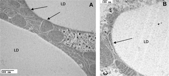
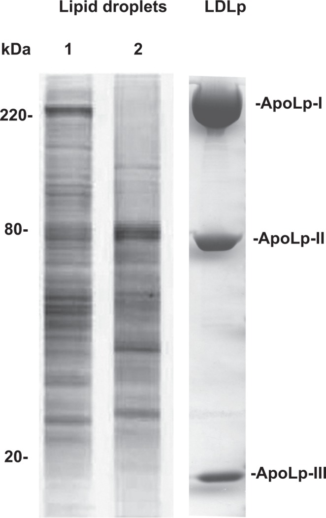
The main cells of the adipose tissue of animals, adipocytes, are characterized by the presence of large cytosolic lipid droplets (LDs), which store triglyceride (TG) and cholesterol. However, most cells have LDs and the ability to store lipids. LDs have a well-known central role in storage and provision of fatty acids and cholesterol. However, the complexity of the regulation of lipid metabolism on the surface of the LDs is still a matter of intense study. Beyond this role, a number of recent studies have suggested that LDs have major functions in other cellular processes, such as protein storage and degradation, and infection and immunity. Thus, our perception of LDs, from simple globules of fat to highly dynamic organelles of unexpected complexity, has been radically transformed. Here we compiled some recent evidence supporting the emerging view that LDs act as platforms connecting a number of relevant metabolic and cellular functions.
Keywords: Lipid droplet; fat body; lipid metabolism; lipoprotein; perilipin; triglycerides.
Figures
References
-
- Digel M, Ehehalt R, Fuellekrug J. Lipid droplets lighting up: insights from live microscopy. FEBS Lett. 2010;584(11):2168–2175. - PubMed
-
- Fujimoto T, Ohsaki Y, Suzuki M, Cheng JL. Imaging lipid droplets by electron microscopy. Methods Cell Biol. 2013;116:227–251. - PubMed
-
- Ohsaki Y, Suzuki M, Fujimoto T. Open questions in lipid droplet biology. Chem Biol. 2014;21(1):86–96. - PubMed
-
- Murphy S, Martin S, Parton RG. Lipid droplet-organelle interactions; sharing the fats. Biochim Biophys Acta. 2009;1791(6):441–447. - PubMed
-
- Targett-Adams P, Chambers D, Gledhill S, et al. Live cell analysis and targeting of the lipid droplet-binding adipocyte differentiation-related protein. J Biol Chem. 2003;278(18):15998–16007. - PubMed
Grants and funding
LinkOut - more resources
Full Text Sources
Other Literature Sources
Miscellaneous
