Review
. 2014 Aug 28:5:409.
doi: 10.3389/fimmu.2014.00409. eCollection 2014.

Nutrient Sensing via mTOR in T Cells Maintains a Tolerogenic Microenvironment

Affiliations
Review

Nutrient Sensing via mTOR in T Cells Maintains a Tolerogenic Microenvironment

Duncan Howie et al. Front Immunol. .

Abstract

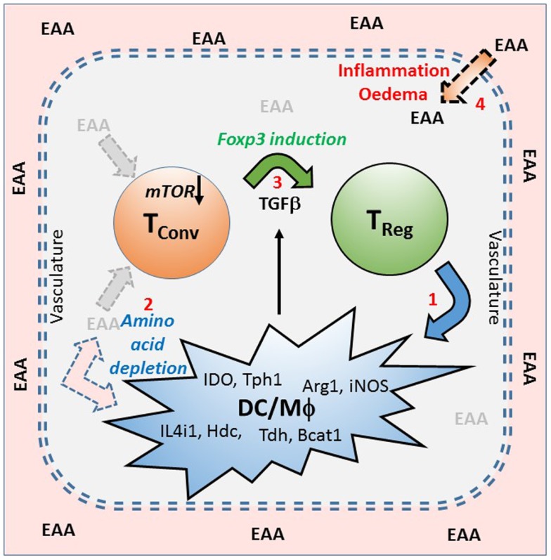
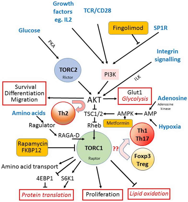
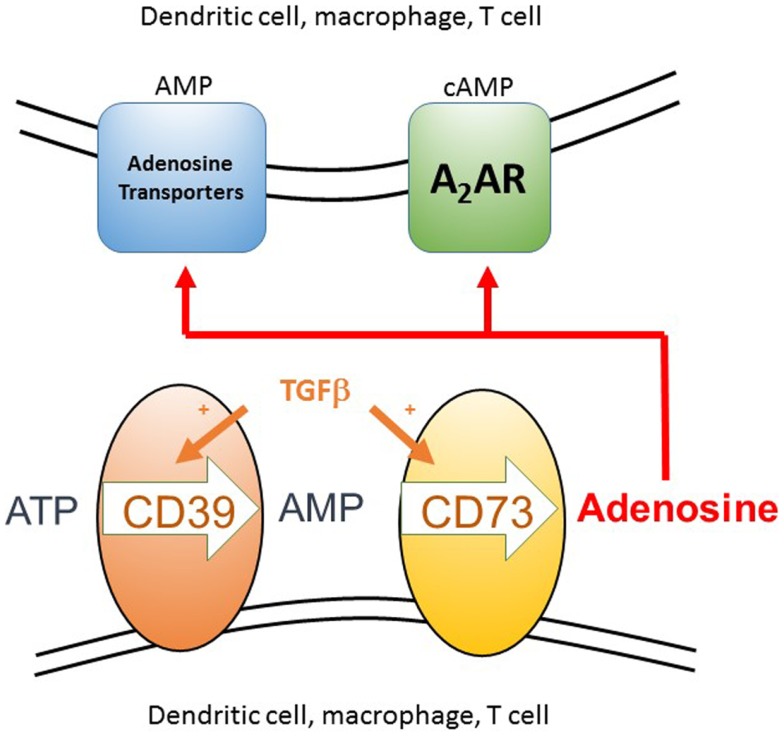
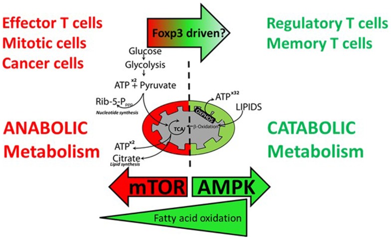
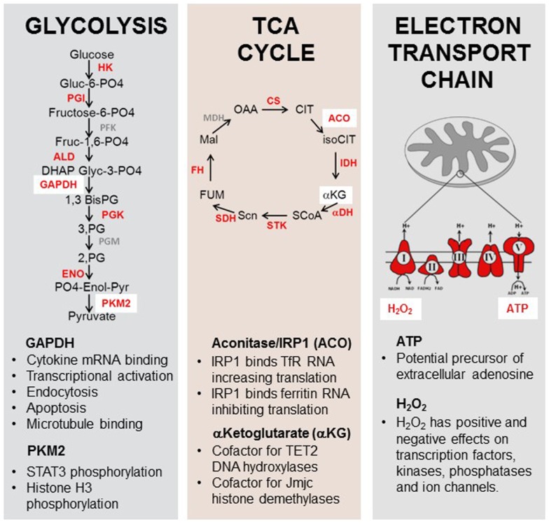
We have proposed that tolerance can be maintained through the induction, by Treg cells, of a tolerogenic microenvironment within tolerated tissues that inhibits effector cell activity but which supports the generation of further Treg cells by "infectious tolerance." Two important components of this tolerogenic microenvironment depend on metabolism and nutrient sensing. The first is due to the up-regulation of multiple enzymes that consume essential amino acids, which are sensed in naïve T cells primarily via inhibition of the mechanistic target of rapamycin (mTOR) pathway, which in turn encourages their further differentiation into FOXP3(+) Treg cells. The second mechanism is the metabolism of extracellular ATP to adenosine by the ectoenzymes CD39 and CD73. These two enzymes are constitutively co-expressed on Treg cells, but can also be induced on a wide variety of cell types by TGFβ and the adenosine generated can be shown to be a potent inhibitor of T cell proliferation. This review will focus on mechanisms of nutrient sensing in T cells, how these are integrated with TCR and cytokine signals via the mTOR pathway, and what impact this has on intracellular metabolism and subsequently the control of differentiation into different effector or regulatory T cell subsets.

Keywords: T cell differentiation; immune regulation; mTOR; metabolism; tolerance.


Figures

Figure 1
Figure 1
A model of infectious tolerance that depends on a nutrient depleted microenvironment maintained by Treg cells within tissues. This model proposes that immunological tolerance is maintained within tissues by the localized depletion of nutrients, particularly the essential amino acids (EAA), which are required for the proliferation and effector function of conventional T cells (Tconv). Amino acid depletion is primarily as a result of regulatory T cells (Treg) inducing (1), in dendritic cells (DC) and macrophages (Mϕ), a range of enzymes that catabolize (2) or utilize EAA (examples are shown). This lack of EAA is sensed via the mTOR pathway which, in the presence of TGFβ, encourages the expression of FOXP3, and the induction of further Treg (3). Under conditions of tolerance the intact vasculature maintains a barrier between the blood and the tissues, but if there is inflammation or damage to the vasculature, causing edema, then EAA can leak into the tissues (4) and contribute to a breaking of the tolerant microenvironment.
Figure 2
Figure 2
The mTOR pathway in T cells. The mechanistic target of rapamycin (mTOR) is a component of both the TORC1 and TORC2 signaling complexes. The TORC1 complex acts as the main integrator of many different signals (input signals shown in blue text) from nutrients such as glucose, via TORC2, the TCR, costimulation and growth factors, via PI3K and AKT, and amino acids via the regulator complex. Hypoxia and AMP levels are also sensed via AMPK and TSC1/2. AKT activation downstream of TORC2 is important for cell survival, drives the expression of the glucose receptor (Glut1) and glycolytic metabolism, and is required for the differentiation into Th2 cells (outputs of signaling shown outlined in red). TORC1 is important for the initiation of mRNA translation via S6K1 mediated phosphorylation of the ribosomal protein S6, and the up-regulation of amino acid transporters at the cell surface. TORC1 also activates lipid oxidation and cell proliferation while it inhibits the expression of FOXP3 and Treg differentiation in favor of Th1 and Th17 cells. The sites where three different clinically available drugs (rapamycin, fingolimod, and metformin) impact on the mTOR pathway are indicated (orange boxes).
Figure 3
Figure 3
The generation of extracellular adenosine as a component of an anti-inflammatory microenvironment. Extracellular ATP arises as a result of cell death, either from the host or pathogen, as can itself be inflammatory. The two ectoenzymes CD39 and CD73 are normally co-expressed constitutively on Treg cells, but can be induced on the surface of many different cell types, including conventional T cells, dendritic cells, and macrophages, in the presence of a source of TGFβ. These enzymes sequentially convert extracellular ATP to AMP and then adenosine. Adenosine can then act either by binding to the A2A receptor on T cells and DC, which signals via cAMP or it can be taken up via adenosine transporters where it is rapidly converted to intracellular AMP by adenosine kinase. These two signaling pathways act to inhibit inflammation and T cell proliferation. AMP activated kinase mediated inhibition of the mTOR pathway can then occur downstream of either signaling pathway.
Figure 4
Figure 4
Effector T cells and regulatory T cells have different requirements for anabolic and catabolic metabolism. Effector T cells, proliferating T cells, and cancer cells favor anabolic metabolism using aerobic glycolysis to fuel energy and cell mass demands during proliferation. Activation of mTOR drives this metabolic shift. Precursors for nucleotide synthesis and lipid synthesis are supplied via the pentose phosphate pathway and the TCA cycle, respectively. Resting regulatory and memory T cells do not require glycolysis and TCA intermediates for cell growth and favor catabolic metabolism fueled by β-oxidation of fatty acids to fuel oxidative phosphorylation. Inhibition of mTOR and activation of AMPK favor a shift to catabolic metabolism.
Figure 5
Figure 5
Moonlighting functions of metabolic enzymes and metabolites. Enzymes and metabolites of the glycolysis, TCA cycle, and electron transport chain play roles in immune function. Enzymes and substrates in red have been shown to have additional non-metabolic functions in eukaryotes [reviewed in Ref. (–176)]. GAPDH can bind to the 3′UTR of some cytokine genes, inhibiting their translation. Pyruvate kinase M2 has been shown to have kinase activity for the pro-inflammatory transcription factor STAT3. Aconitase functions as a rheostat for cellular iron in addition to its role in the TCA cycle. α-Ketoglutarate is an essential cofactor for the enzymes TET2 and jumonji-C histone demethylases. ATP may act as a precursor for extracellular adenosine production and H2O2 has been shown to possess a signaling role in multiple cytoplasmic and nuclear pathways.

References

    1. Fang Y, Vilella-Bach M, Bachmann R, Flanigan A, Chen J. Phosphatidic acid-mediated mitogenic activation of mTOR signaling. Science (2001) 294:1942–510.1126/science.1066015 - DOI - PubMed
    1. Peter C, Waldmann H, Cobbold SP. mTOR signalling and metabolic regulation of T cell differentiation. Curr Opin Immunol (2010) 22:655–6110.1016/j.coi.2010.08.010 - DOI - PubMed
    1. Warburg O. On respiratory impairment in cancer cells. Science (1956) 124:269–70 - PubMed
    1. Buttgereit F, Brand MD, Muller M. ConA induced changes in energy metabolism of rat thymocytes. Biosci Rep (1992) 12:381–610.1007/BF02351215 - DOI - PubMed
    1. Caro-Maldonado A, Gerriets VA, Rathmell JC. Matched and mismatched metabolic fuels in lymphocyte function. Semin Immunol (2012) 24:405–1310.1016/j.smim.2012.12.002 - DOI - PMC - PubMed