

Intracranial complication of rhinosinusitis from actinomycosis of the paranasal sinuses: a rare case of abducens nerve palsy
- PMID: 25221679
- PMCID: PMC4158282
- DOI: 10.1155/2014/601671
Intracranial complication of rhinosinusitis from actinomycosis of the paranasal sinuses: a rare case of abducens nerve palsy
Abstract
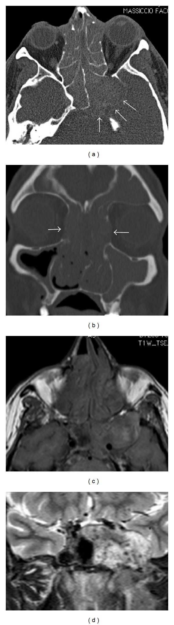
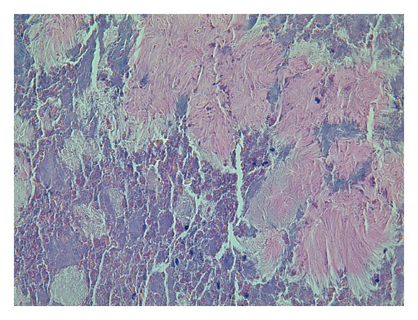
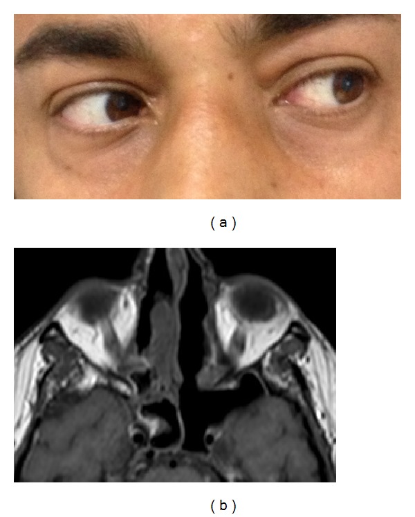
Sinonasal actinomycosis should be suspected when a patient with chronic sinusitis does not respond to medical therapy or has a history of facial trauma, dental disease, cancer, immunodeficiency, long-term steroid therapy, diabetes, or malnutrition. Radiological evaluation with computed tomography and magnetic resonance imaging are important in differential diagnosis, evaluating the extent of disease, and understanding clinical symptoms. Endoscopic sinus surgery associated with long-term intravenous antibiotic therapy is the gold standard for treatment of sinonasal actinomycosis. We report an unusual case of abducens nerve palsy resulting from invasive sinonasal actinomycosis in a patient with an abnormally enlarged sphenoid sinus. A review of the current literature highlighting clinical presentation, radiological findings, and treatment of this uncommon complication is also presented.
Figures

References
-
- Roth M, Montone KT. Actinomycosis of the paranasal sinuses: a case report and review. Otolaryngology—Head and Neck Surgery. 1996;114(6):818–821. - PubMed
-
- Miller M, Haddad AJ. Cervicofacial actinomycosis. Oral Surgery, Oral Medicine, Oral Pathology, Oral Radiology, and Endodontics. 1998;85(5):496–508. - PubMed
-
- Pulverer G, Schütt-Gerowitt H, Schaal KP. Human cervicofacial actinomycoses: microbiological data for 1997 cases. Clinical Infectious Diseases. 2003;37(4):490–497. - PubMed
-
- Bennhoff DF. Actinomycosis: diagnostic and therapeutic considerations and a review of 32 cases. Laryngoscope. 1984;94(9):1198–1217. - PubMed
-
- Wadhera R, Gulati SP, Garg A, Ghai A, Kumar S. Frontal sinus actinomycosis presenting as osteomyelitis of frontal bone. Otolaryngology-Head and Neck Surgery. 2008;138(4):544–545. - PubMed
LinkOut - more resources
Full Text Sources
Other Literature Sources
Molecular Biology Databases
