

Incidental benign metastasizing leiomyoma in a patient with bone sarcoma: a case report
- PMID: 25221682
- PMCID: PMC4158327
- DOI: 10.1155/2014/439061
Incidental benign metastasizing leiomyoma in a patient with bone sarcoma: a case report
Abstract
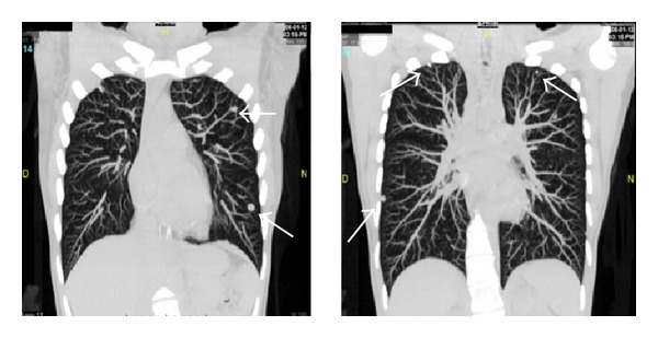
Background. The benign metastasizing leiomyoma is an exceptionally rare entity; it presents with ectopic leiomyoma nodules with a benign pattern. Symptoms vary according to the anatomic location. The diagnosis is histopathological, usually in patients with history of hysterectomy. Case Presentation. A 36-year-old female with 2-month history of left knee pain was diagnosed with bone fibrosarcoma. A CT scan showed pulmonary nodules. The patient started neoadjuvant chemotherapy. Conservative surgery of pelvic limb was achieved. A new CT scan reported pulmonary nodules that remained in relation to the previous CT. A nodule resection by thoracotomy and TOB (transoperative biopsy) was performed. The final pathology report described benign proliferative lesions consistent with benign metastatic leiomyoma. Conclusions. Benign metastatic leiomyoma is a rare condition presenting with uterine and extrauterine nodules most commonly in the lung. The diagnosis is histopathological. The surgical procedure must be reserved for selected patients.
Figures


References
-
- Jautzke G, Müller-Ruchholtz E, Thalmann U. Immunohistological detection of estrogen and progesterone receptors in multiple and well differentiated leiomyomatous lung tumors in women with uterine leiomyomas (so-called benign metastasizing leiomyomas): a report on 5 cases. Pathology Research and Practice. 1996;192(3):215–223. - PubMed
-
- Egberts J, Schafmayer C, Bauerschlag DO, Jänig U, Tepel J. Benign abdominal and pulmonary metastasizing leiomyoma of the uterus. Archives of Gynecology and Obstetrics. 2006;274(5):319–322. - PubMed
-
- Patton KT, Cheng L, Papavero V, et al. Benign metastasizing leiomyoma: clonality, telomere length and clinicopathologic analysis. Modern Pathology. 2006;19(1):130–140. - PubMed
-
- Awonuga AO, Shavell VI, Imudia AN, Rotas M, Diamond MP, Puscheck EE. Pathogenesis of benign metastasizing leiomyoma: a review. Obstetrical and Gynecological Survey. 2010;65(3):189–195. - PubMed
LinkOut - more resources
Full Text Sources
Other Literature Sources
