Review
. 2014 Oct;15(10):1036-52.
doi: 10.15252/embr.201439225. Epub 2014 Sep 15.

Cholesterol in brain disease: sometimes determinant and frequently implicated

Affiliations
Review

Cholesterol in brain disease: sometimes determinant and frequently implicated

Mauricio G Martín et al. EMBO Rep. 2014 Oct.

Abstract

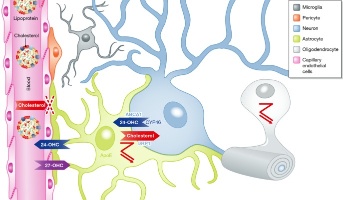
Cholesterol is essential for neuronal physiology, both during development and in the adult life: as a major component of cell membranes and precursor of steroid hormones, it contributes to the regulation of ion permeability, cell shape, cell-cell interaction, and transmembrane signaling. Consistently, hereditary diseases with mutations in cholesterol-related genes result in impaired brain function during early life. In addition, defects in brain cholesterol metabolism may contribute to neurological syndromes, such as Alzheimer's disease (AD), Huntington's disease (HD), and Parkinson's disease (PD), and even to the cognitive deficits typical of the old age. In these cases, brain cholesterol defects may be secondary to disease-causing elements and contribute to the functional deficits by altering synaptic functions. In the first part of this review, we will describe hereditary and non-hereditary causes of cholesterol dyshomeostasis and the relationship to brain diseases. In the second part, we will focus on the mechanisms by which perturbation of cholesterol metabolism can affect synaptic function.

Keywords: brain disease; cholesterol metabolism; cognition.


Figures

Figure 1
Figure 1. Cellular cholesterol homeostasis
Diagram summarizing how cells ensure cholesterol homeostasis. Cells synthesize cholesterol from acetyl-CoA by a long series of enzymatic steps requiring energy and molecular oxygen. Intermediates of the pathway serve as precursors for other biologically active molecules. Highlighted enzymes are 3-hydroxy-3-methyl-glutaryl-CoA reductase (HMGCR), which is rate-limiting for the mevalonate pathway and inhibited by statins, 24-dehydrocholesterol reductase (DHCR24) and 7-dehydrocholesteol reductase (DHCR7), whose defects cause rare human diseases. Cells can take up cholesterol by receptor-mediated endocytosis of lipoproteins bearing apolipoproteins. In this pathway, Niemann-Pick Type C Protein 1 and NPC2 mediate cooperatively the exit of cholesterol out of the endosomal-lysosomal system and thereby allow for its incorporation into the intracellular pool. Defects in either protein cause the lysosomal storage disorder Niemann-Pick Type C. Overload by cholesterol is prevented by its intracellular esterification and subsequent storage in lipid droplets and by its release. Cholesterol is released either as a complex with apolipoprotein-containing lipoproteins via members of the ATP-binding cassette transporters or after conversion to oxysterols. Examplary proteins for each process are indicated. The relative contribution of each pathway to cholesterol homeostasis is probably cell type-specific. The post-lanosterol steps of cholesterol biosynthesis are divided into Bloch and Kandutsch–Russell pathways, which share enzymatic stages but produce C24 double-bond reduced cholesterol at different steps.
Figure 2
Figure 2. Brain-specific aspects of cholesterol metabolism
Brain cells are cut off from blood supply as the blood–brain barrier prevents entry of lipoproteins. Cholesterol is synthesized by different types of glial cells as indicated by the zagged pathway symbol. Some neurons may import cholesterol from APOE-expressing astrocytes via lipoprotein uptake (LRP1) and hydroxylate surplus cholesterol to 24-OHC, which is excreted via ABCA1 and which enters the blood circulation. 24-OHC is able to cross the blood–brain barrier where it may signal the level of plasma cholesterol levels to cells in the brain. Cholesterol-related proteins with cell-specific distribution are indicated.
Figure 3
Figure 3. Schematic picture showing how AD can lead to reduced cholesterol in neurons
Numerous studies in brain samples from AD affected individuals show reduced cholesterol levels in structures like the hippocampus. In vitro studies suggest that amyloid oligomers, whether through binding to detergent resistant membrane domains (rafts) and/or in the form of amyloid peptide aggregates in intracellular compartments, trigger cell cholesterol decrease by various mechanisms: stress-activated Cyp46A1 transcription leading to cholesterol solubilization and excretion in the form of 24-OHC; sequestration of membranes and cholesterol within terminals from dying neurons and directly in amyloid plaques; inhibition of astrocyte-derived APOE-cholesterol uptake and/or by direct changes in plasma membrane lipid content. Cholesterol reduction can also occur in conditions accompanied by an excess of APP through direct inhibition of HMG-CoA reductase (HMGCR) and SREBP mRNA levels.
Figure 4
Figure 4. Proposed mechanism to explain how reduced brain cholesterol could underlie poor cognition
In the presynaptic compartment, cholesterol depletion impairs synaptic vesicle exocytosis probably due to altered membrane curvature and impaired SNARE clusterization at fusion-competent sites. Cholesterol depletion also affects the ability of synapses to undergo sustained synaptic transmission by compromising the recycling of proteins from synaptic vesicles. In the postsynaptic compartment, cholesterol loss leads to MARCKS detachment from the membrane and PIP2 release. Most of the released PIP2 is transformed to PIP3 due to the enhanced TrkB PI3K activity also promoted by cholesterol loss. PIP3 accumulation stabilizes F-actin, blocks AMPARs at the dendritic spines, and also leads to high levels of active p-Akt, which in turn inactivates GSK3β required to promote AMPARs endocytosis. As a consequence of reduced PIP2, low PLCγ activity is also found in old neurons leading to impaired LTP. PLC activity is also required for processes such as actin depolymerization, AKAP150 removal from spines, and PSD95 degradation after LTD induction. Altogether, impaired LTP and LTD in old neurons result in decreased learning and memory.

References

    1. Mañes S, Martínez-A C. Cholesterol domains regulate the actin cytoskeleton at the leading edge of moving cells. Trends Cell Biol. 2004;14:275–278. - PubMed
    1. Dart C. Lipid microdomains and the regulation of ion channel function. J Physiol. 2010;588:3169–3178. - PMC - PubMed
    1. Lippincott-Schwartz J, Phair RD. Lipids and cholesterol as regulators of traffic in the endomembrane system. Annu Rev Biophys. 2010;39:559–578. - PMC - PubMed
    1. Simons K, Gerl MJ. Revitalizing membrane rafts: new tools and insights. Nat Rev Mol Cell Biol. 2010;10:688–699. - PubMed
    1. Levitan I, Singh DK, Rosenhouse-Dantsker A. Cholesterol binding to ion channels. Front Physiol. 2014;5:65. - PMC - PubMed

MeSH terms