

The accuracy of a designed software for automated localization of craniofacial landmarks on CBCT images
- PMID: 25223399
- PMCID: PMC4171715
- DOI: 10.1186/1471-2342-14-32
The accuracy of a designed software for automated localization of craniofacial landmarks on CBCT images
Abstract
Background: Two-dimensional projection radiographs have been traditionally considered the modality of choice for cephalometric analysis. To overcome the shortcomings of two-dimensional images, three-dimensional computed tomography (CT) has been used to evaluate craniofacial structures. However, manual landmark detection depends on medical expertise, and the process is time-consuming. The present study was designed to produce software capable of automated localization of craniofacial landmarks on cone beam (CB) CT images based on image registration and to evaluate its accuracy.
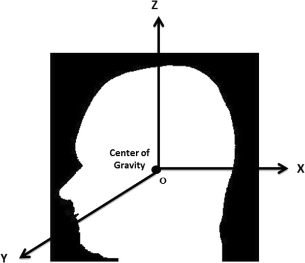
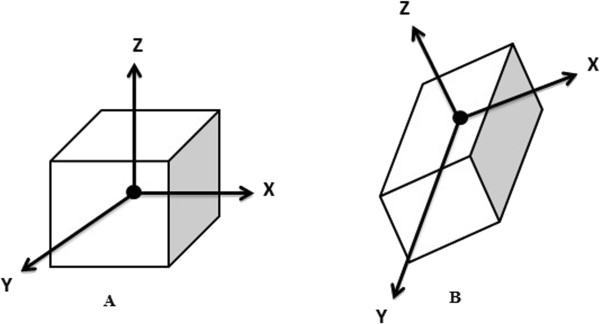
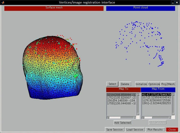
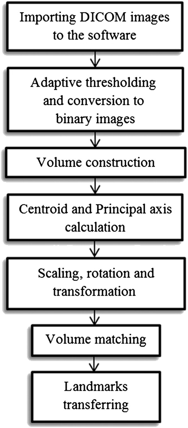
Methods: The software was designed using MATLAB programming language. The technique was a combination of feature-based (principal axes registration) and voxel similarity-based methods for image registration. A total of 8 CBCT images were selected as our reference images for creating a head atlas. Then, 20 CBCT images were randomly selected as the test images for evaluating the method. Three experts twice located 14 landmarks in all 28 CBCT images during two examinations set 6 weeks apart. The differences in the distances of coordinates of each landmark on each image between manual and automated detection methods were calculated and reported as mean errors.
Results: The combined intraclass correlation coefficient for intraobserver reliability was 0.89 and for interobserver reliability 0.87 (95% confidence interval, 0.82 to 0.93). The mean errors of all 14 landmarks were <4 mm. Additionally, 63.57% of landmarks had a mean error of <3 mm compared with manual detection (gold standard method).
Conclusion: The accuracy of our approach for automated localization of craniofacial landmarks, which was based on combining feature-based and voxel similarity-based methods for image registration, was acceptable. Nevertheless we recommend repetition of this study using other techniques, such as intensity-based methods.
Figures

References
-
- Forsyth DB, Shaw WC, Richmond S, Roberts CT. Digital imaging of cephalometric radiographs, Part 2: Image quality. Angle Orthod. 1996;66:43–50. - PubMed
-
- Makram M, Kamel H. Reeb Graph for Automatic 3D Cephalometry. IJIP. 2014;8:17–29.
Publication types
MeSH terms
LinkOut - more resources
Full Text Sources
Other Literature Sources
