. 2014 Sep 16;18(5):511.
doi: 10.1186/s13054-014-0511-3.

Endothelial dysfunction is a potential contributor to multiple organ failure and mortality in aged mice subjected to septic shock: preclinical studies in a murine model of cecal ligation and puncture

Endothelial dysfunction is a potential contributor to multiple organ failure and mortality in aged mice subjected to septic shock: preclinical studies in a murine model of cecal ligation and puncture

Ciro Coletta et al. Crit Care. .

Abstract

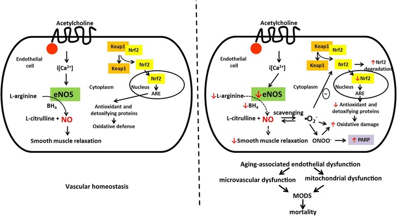
Introduction: The goal of the current study was to investigate the effect of aging on the development of endothelial dysfunction in a murine model of sepsis, and to compare it with the effect of genetic deficiency of the endothelial isoform of nitric oxide synthase (eNOS).

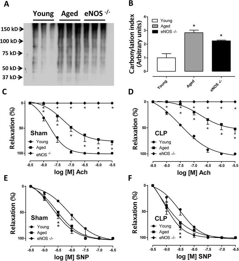
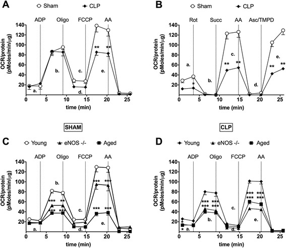
Methods: Cecal ligation and puncture (CLP) was used to induce sepsis in mice. Survival rates were monitored and plasma indices of organ function were measured. Ex vivo studies included the measurement of vascular function in thoracic aortic rings, assessment of oxidative stress/cellular injury in various organs and the measurement of mitochondrial function in isolated liver mitochondria.

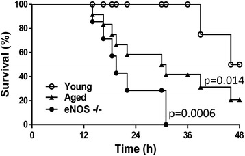
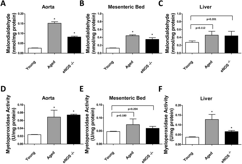
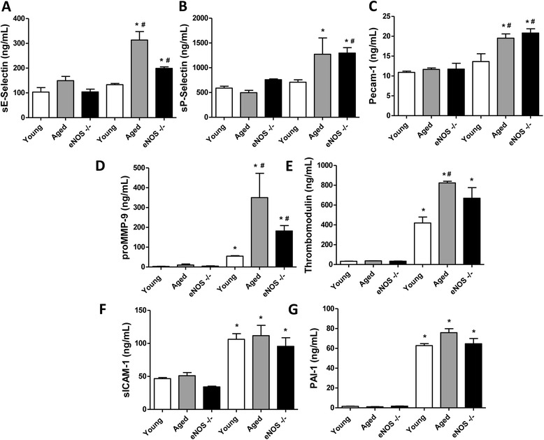
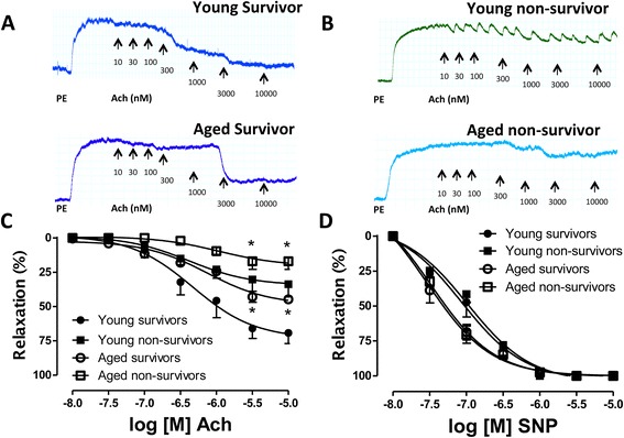
Results: eNOS deficiency and aging both exacerbated the mortality of sepsis. Both eNOS-deficient and aged mice exhibited a higher degree of sepsis-associated multiple organ dysfunction syndrome (MODS), infiltration of tissues with mononuclear cells and oxidative stress. A high degree of sepsis-induced vascular oxidative damage and endothelial dysfunction (evidenced by functional assays and multiple plasma markers of endothelial dysfunction) was detected in aortae isolated from both eNOS(-/-) and aged mice. There was a significant worsening of sepsis-induced mitochondrial dysfunction, both in eNOS-deficient mice and in aged mice. Comparison of the surviving and non-surviving groups of animals indicated that the severity of endothelial dysfunction may be a predictor of mortality of mice subjected to CLP-induced sepsis.

Conclusions: Based on the studies in eNOS mice, we conclude that the lack of endothelial nitric oxide production, on its own, may be sufficient to markedly exacerbate the severity of septic shock. Aging markedly worsens the degree of endothelial dysfunction in sepsis, yielding a significant worsening of the overall outcome. Thus, endothelial dysfunction may constitute an early predictor and independent contributor to sepsis-associated MODS and mortality in aged mice.


Figures

Figure 1
Figure 1
Time course of survival rates in young, aged and eNOS −/− mice subjected to CLP. Mice were subjected to acute sepsis by CLP as described in Methods and constantly monitored for 48 hours. Death events were annotated and the Kaplan-Meier estimate was used to compute the survival rates over time. Please notice that by 24 hours the eNOS-deficient mice showed the highest mortality rate (about 70%). At this time there was 40% mortality in the aged mice while no mortality in the wild-type control group (young mice). By 24 to 36 hours all the eNOS−/− mice died, the mortality of the aged mice increased to about 60% while all the young mice survived. By 48 hours, the survival rates of the aged mice and the young mice were 20% and 50% respectively. Sham-operated mice showed no mortality in the 48-hour time frame (not shown) (P = 0.008 eNOS−/− vs. young and P = 0.024 aged vs. young; n = 12/group). CLP, cecal ligation and puncture; eNOS, endothelial isoform of nitric oxide synthase.
Figure 2
Figure 2
Determination of lipid peroxidation and myeloperoxidase activity in aorta, mesenteric bed and liver harvested from CLP-injured mice. Tissue malondialdehyde (MDA) levels (index of cellular injury/oxidative stress) and myeloperoxidase (MPO) activity (used as a marker of tissue inflammation) were detected in aorta (Panel A and D), mesenteric vascular bed (Panel B and E) and liver (Panel C and F) harvested from young, aged and eNOS−/− mice subjected to 12 hours of CLP-induced sepsis (* P <0.05 vs. young; n = 5 tissue samples were analyzed in each group of mice). CLP, cecal ligation and puncture; eNOS, endothelial isoform of nitric oxide synthase.
Figure 3
Figure 3
Detection of plasma markers of cardiovascular dysfunction (CVD panel) in sham-operated or CLP-injured mice. Plasma levels of sE-Selectin (Panel A), sP-Selectin (Panel B), Pecam-1 (Panel C), proMMP-9 (Panel D), thrombomodulin (Panel E), sICAM-1 (Panel F), and PAI-1 Total (Panel G), were simultaneously detected in young, aged or eNOS−/− mice 12 hours following sham (left three columns in each panel) or CLP (right three columns in each panel) procedure. (* P <0.05 vs. corresponding sham-operated mice and # P <0.05 vs. young CLP; n = 5 individual plasma samples were tested from each group of mice). CLP, cecal ligation and puncture; eNOS, endothelial isoform of nitric oxide synthase.
Figure 4
Figure 4
Assessment of oxidative damage and endothelial dysfunction in aortas isolated from septic mice. (Panel A) Following CLP, young, aged and eNOS−/− mice were euthanized by opening the chest cavity under deep anesthesia and the thoracic aortas were isolated. Twenty μg of protein extracts were subjected to SDS-PAGE and derivatized protein carbonyl groups were immunodetected by OxyBlot (Merck Millipore). Immunoreactivity was visualized by chemiluminescent detection. Blots were scanned and a densitometric analysis was performed (Panel B) (* P <0.05 vs. young mice; n = 3). In a separate set of experiments, aortic rings (approximately 2 mm in length) were harvested from sham-operated and CLP-injured mice, placed in 5 ml organ baths filled with oxygenated (95% O2 to 5% CO2) Krebs-Henseleit solution at 37°C and mounted onto isometric force transducers. Concentration response curves to acetylcholine (Ach) (Panel C and D) and sodium nitroprusside (SNP) (Panel D and E) were performed on a stable phenylephrine-induced tone. Please notice the inability of the aortic rings from eNOS−/− mice to respond to Ach stimulation. Aged mice showed a basal level of endothelial dysfunction (Panel C) (* P <0.05 vs. young). CLP induced a shift to the right of the concentration response curve to Ach, which was more pronounced in the aged mice compared to the young (Panel D) (mean LogEC50 from −7.797 to −7.488 in young mice; mean LogEC50 from −7.261 to −6.762 in aged mice). Increased sensitivity to the nitric oxide (NO) donor SNP was also seen in aged and eNOS-deficient vessels (Panel E and F). CLP, cecal ligation and puncture; eNOS, endothelial isoform of nitric oxide synthase.
Figure 5
Figure 5
Assessment of endothelial function in mice that survived or did not survive 48 hours of sepsis. CLP was performed in C57BL/6 young mice as described in Methods. Following CLP, mice were constantly monitored for 48 hours. In our model the survival rate at 48 hours is approximately 50%. Thoracic aortas from the mice that did not survive the 48 hours of sepsis were harvested at the exact time of death, placed in 5 ml organ baths filled with oxygenated Krebs-Henseleit solution at 37°C and mounted onto isometric force transducers to assess for vascular response to endothelium-derived nitric oxide (NO) by acetylcholine (Ach) stimulation. Five death events were recorded, aortic rings from the five non-survivors were used. The mean time of survival (or tissue harvesting) was 39 hours. Survivors were euthanized at 48 hours of sepsis by opening the chest cavity. In a sequent study, a similar approach was used for the assessment and comparison of endothelial function in survivor versus non-survivor aged mice. Since the survival rate at 48 hours of CLP is only 80% in the case of the aged mice, we restricted the CLP to a 24-hour time frame. Aortic rings were harvested from five different mice. The mean time of survival (or tissue harvesting) in this group was 17 hours. The survivors were euthanized 24 hours following CLP. Please note the marked degree of endothelial dysfunction in the non-survivor group of animals (Panel A, B and C). Concentration response curves to the NO donor sodium nitroprusside (SNP) were performed to ensure the integrity of the smooth muscle and the viability of the tissue preparations (Panel D). (* P <0.05 vs. corresponding survivors). CLP, cecal ligation and puncture.
Figure 6
Figure 6
Bioenergetic analysis in isolated liver mitochondria from mice subjected to sham or CLP operation. Mitochondrial respiration/function was determined by oxygen consumption rate (OCR) utilizing the Seahorse XF24 Extracellular Flux Analyzer (Seahorse Bioscience). During the coupling experiment in isolated mitochondria (A, C, D), the assay media contains succinate, as a complex II substrate and rotenone as a complex I inhibitor. Sequential measurement of basal, State 2 (a), State 3 (b), State 4o (c) and the uncoupler-stimulated respiration, State 3u (d) were performed through the sequential injections of ADP, oligomycin, FCCP and antimycin. Please note that the OCR after the addition of FCCP (State 3u) is much higher in sham group (A). Furthermore, a significant impairment in State 3 and State 3u respirations has been detected in aged and eNOS−/− group compared to young mice subjected to CLP intervention or the sham group (C, D). (Panel A, C, D, ** P <0.01 and *** P <0.001 vs. sham or CLP-operated young mice; n = at least 4/group). The experiment in (panel B) demonstrates the electron flow activity through the different complexes of the electron transport chain. The assay media contains pyruvate/malate as substrates of complex I, and FCCP to uncouple the mitochondrial function. In uncoupled mitochondria all complexes can be examined individually by the sequential injection of rotenone, succinate, antimycin and TMPD/ascorbate. All these mitochondrial toxins and substrates permit to observe 1., the function of complex I (a), 2., the inhibition of complex I (b), 3., the function of complex II (c), 4., the inhibition of complex II (d), 5., the activity of cytochrome C/complex IV (e). Overall, all mitochondrial complexes show better function in mitochondria isolated from the liver of the sham group compared to the other groups (Panel B, ** P <0.01 vs. sham-operated mice; n = at least 4/group). ADP, adenosine 5′-diphosphate sodium salt; CLP, cecal ligation and puncture; eNOS, endothelial isoform of nitric oxide synthase; FCCP, carbonylcyanide-4-trifluorometh-oxyphenylhydrazone; TMPD, tetra-methyl-p-phenylenediamine dihydrochloride.
Figure 7
Figure 7
Potential pathways and mechanisms underlying the enhancement of endothelial dysfunction in aged mice subjected to sepsis. In aged blood vessels, septic shock induces an overwhelming degree of oxidative damage, leading to a marked degree of endothelial dysfunction, which, in turn, contributes to the MODS and mortality of sepsis. We hypothesize that part of the process involves the fact that in aged endothelial cell, oxidative stress is unable to induce an appropriate activation of the Nrf2/ARE pathway. Please see Discussion for additional details. ARE, antioxidant response element; BH4, tetrahydrobiopterin, a co-factor of eNOS; eNOS, endothelial isoform of NO synthase; iCa2+, intracellular calcium concentration; Keap1, Kelch-like erythroid cell-derived protein with CNC homology (ECH)-associated protein 1; MODS, multiple organ dysfunction syndrome; NO, nitric oxide; Nrf2, nuclear factor erythroid 2 (NF-E2)-related factor 2; PARP, poly(ADP-ribose) polymerase.

References

    1. Martin GS, Mannino DM, Eaton S, Moss M. The epidemiology of sepsis in the United States from 1979 through 2000. N Engl J Med. 2001;348:1546–1554. doi: 10.1056/NEJMoa022139. - DOI - PubMed
    1. Martin GS, Mannino DM, Moss M. The effect of age on the development and outcome of adult sepsis. Crit Care Med. 2006;34:15–21. doi: 10.1097/01.CCM.0000194535.82812.BA. - DOI - PubMed
    1. Altura BM, Gebrewold A, Burton RW. Failure of microscopic metarterioles to elicit vasodilator responses to acetylcholine, bradykinin, histamine and substance P after ischemic shock, endotoxemia and trauma: possible role of endothelial cells. Microcirc Endothelium Lymphatics. 1985;2:121–127. - PubMed
    1. Szabo C, Farago M, Horvath I, Lohinai Z, Kovach AG. Hemorrhagic hypotension impairs endothelium-dependent relaxations in the renal artery of the cat. Circ Shock. 1992;36:238–241. - PubMed
    1. Wang P, Ba ZF, Chaudry IH. Endothelial cell dysfunction occurs after hemorrhage in nonheparinized but not in preheparinized models. J Surg Res. 1993;54:499–506. doi: 10.1006/jsre.1993.1077. - DOI - PubMed

Publication types

MeSH terms