Review
. 2015 Jan:37:108-18.
doi: 10.1016/j.semcdb.2014.09.004. Epub 2014 Sep 16.

Regulation of development and cancer by the R2B subfamily of RPTPs and the implications of proteolysis

Affiliations
Review

Regulation of development and cancer by the R2B subfamily of RPTPs and the implications of proteolysis

Sonya E L Craig et al. Semin Cell Dev Biol. 2015 Jan.

Abstract

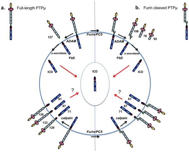
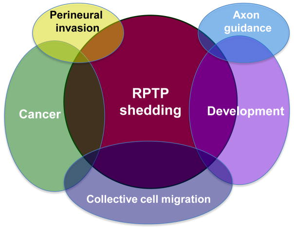
The initial cloning of receptor protein tyrosine phosphatases (RPTPs) was met with excitement because of their hypothesized function in counterbalancing receptor tyrosine kinase signaling. In recent years, members of a subfamily of RPTPs with homophilic cell-cell adhesion capabilities, known as the R2B subfamily, have been shown to have functions beyond that of counteracting tyrosine kinase activity, by independently influencing cell signaling in their own right and by regulating cell adhesion. The R2B subfamily is composed of four members: PTPmu (PTPRM), PTPrho (PTPRT), PTPkappa (PTPRK), and PCP-2 (PTPRU). The effects of this small subfamily of RPTPs is far reaching, influencing several developmental processes and cancer. In fact, R2B RPTPs are predicted to be tumor suppressors and are among the most frequently mutated protein tyrosine phosphatases (PTPs) in cancer. Confounding these conclusions are more recent studies suggesting that proteolysis of the full-length R2B RPTPs result in oncogenic extracellular and intracellular protein fragments. This review discusses the current knowledge of the role of R2B RPTPs in development and cancer, with special detail given to the mechanisms and implications that proteolysis has on R2B RPTP function. We also touch upon the concept of exploiting R2B proteolysis to develop cancer imaging tools, and consider the effects of R2B proteolysis on axon guidance, perineural invasion and collective cell migration.

Keywords: Axon guidance; Collective cell migration; Perineural invasion; Proteolysis; RPTP; Receptor PTP.


Figures

Figure 1
Figure 1
PTPμ proteolysis in cancer cells as a model for R2B proteolysis. PTPμ, PTPκ, and PCP-2 have all been shown to be proteolyzed in cancer cells, and may follow the same steps as has been observed for PTPμ proteolysis. The molecular weights shown are for PTPμ. PTPμ exists at the cell surface in two full-length forms: either the non-furin processed full-length PTPμ protein or the furin-processed form that consists of two non-covalently associated PTPμ subunits (furin cleaved PTPμ). In (a), ADAM protease cuts full-length PTPμ in the extracellular domain to yield a shed 127 kDa fragment and a membrane-tethered PΔE fragment. The PΔE fragment is then proteolyzed by the γ-secretase complex to release the ICD fragment from the plasma membrane. The ICD is capable of translocating into the cell nucleus. PTPμ is proteolyzed differently in cancer cells in response to ionomycin stimulation. Full-length PTPμ is also cleaved by calpain inside of the cell, to yield three membrane-free fragments of 71, 67, and 61 kDa and three integral membrane forms of PTPμ with intact extracellular domains of 129, 133, and 139 kDa. In (b), furin cleaves PTPμ in the golgi network to yield two non-covalently associated subunits at the plasma membrane. Furin-processed forms of PTPμ are also cleaved in the extracellular domain by ADAM to yield shed fragments of 108 kDa. The membrane-tethered fragment is then cleaved by the γ-secretase complex to yield the ICD fragment. In cancer cells, calpain cleavage of furin processed PTPμ yields membrane-free cytoplasmic fragments of 71, 67, and 61 kDa, as is observed for full-length PTPμ (a). Three integral membrane forms of PTPμ with intact extracellular domains are also generated in response to calpain cleavage. PTPκ is proteolyzed by a furin-like protease, an ADAM, and the γ-secretase complex, just like PTPμ.
Figure 2
Figure 2
RPTP proteolysis affects both development and cancer, occasionally by influencing the same cellular mechanism. For instance, proteolysis of proteins involved in collective cell migration is hypothesized to affect migration during development and in cancer. Proteolysis has also been shown to influence the neural developmental process of axon guidance. Finally, perineural invasion may be influenced by receptor proteolysis in cancer. Because the R2B RPTPs are implicated in many of these processes, their proteolysis is likely to have far reaching cellular implications in development and cancer.

References

    1. Tonks NK. Protein tyrosine phosphatases--from housekeeping enzymes to master regulators of signal transduction. FEBS J. 2013;280:346–78. - PMC - PubMed
    1. Brady-Kalnay SM, Tonks NK. Protein tyrosine phosphatases as adhesion receptors. Curr Opin Cell Biol. 1995;7:650–7. - PubMed
    1. Andersen JN, Mortensen OH, Peters GH, Drake PG, Iversen LF, Olsen OH, et al. Structural and evolutionary relationships among protein tyrosine phosphatase domains. Mol Cell Biol. 2001;21:7117–36. - PMC - PubMed
    1. Brady-Kalnay S. Ig-superfamily phosphatases. In: Sonderegger P, editor. Ig Superfamily Molecules in the Nervous System. Zurich: Harwood Academic Publishers; 1998.
    1. Tonks NK, Yang Q, Flint AJ, Gebbink MF, Franza BR, Jr, Hill DE, et al. Protein tyrosine phosphatases: the problems of a growing family. Cold Spring Harb Symp Quant Biol. 1992;57:87–94. - PubMed

Publication types

Substances

LinkOut - more resources