. 2014 Oct;42(18):11304-20.
doi: 10.1093/nar/gku809. Epub 2014 Sep 15.

Unraveling the sequence-dependent polymorphic behavior of d(CpG) steps in B-DNA

Affiliations

Unraveling the sequence-dependent polymorphic behavior of d(CpG) steps in B-DNA

Pablo Daniel Dans et al. Nucleic Acids Res. 2014 Oct.

Abstract

We have made a detailed study of one of the most surprising sources of polymorphism in B-DNA: the high twist/low twist (HT/LT) conformational change in the d(CpG) base pair step. Using extensive computations, complemented with database analysis, we were able to characterize the twist polymorphism in the d(CpG) step in all the possible tetranucleotide environment. We found that twist polymorphism is coupled with BI/BII transitions, and, quite surprisingly, with slide polymorphism in the neighboring step. Unexpectedly, the penetration of cations into the minor groove of the d(CpG) step seems to be the key element in promoting twist transitions. The tetranucleotide environment also plays an important role in the sequence-dependent d(CpG) polymorphism. In this connection, we have detected a previously unexplored intramolecular C-H···O hydrogen bond interaction that stabilizes the low twist state when 3'-purines flank the d(CpG) step. This work explains a coupled mechanism involving several apparently uncorrelated conformational transitions that has only been partially inferred by earlier experimental or theoretical studies. Our results provide a complete description of twist polymorphism in d(CpG) steps and a detailed picture of the molecular choreography associated with this conformational change.


Figures

Figure 1.
Figure 1.
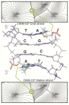
Representation of the ζ states under the BI or BII conformations. Unless otherwise stated, during this work we consider the coupling between the twist at the CG step and the two ζ angles (one in each strand) located at the 3′-junction of the step (highlighted in red in the bottom representation). Considering two ζ angles gives four possible combinations: (i) Both strands are in g-/g-, (ii) the Watson strand is in g- and the Crick strand in t (g-/t), (iii) the inverse situation (t,g-), and (iv) both strands are in t/t.
Figure 2.
Figure 2.
Twist distributions for the central CG step (black) and normal components obtained with BIC (LT component in red, HT component in green) for the 10 possible tetranucleotides.
Figure 3.
Figure 3.
Correlations between twist at the central CG step and the states of the ζ angle at the 3′-side for K+Cl.
Figure 4.
Figure 4.
Two-dimensional cation distributions averaged over the last 200 ns of the trajectories. The plot show the radial-angular plane at the central CG step, the minor groove limits as white lines and the center of the major groove as a vertical radial vector. The results are plotted as molarities as shown by the color bars, with a blue to red concentration scale that goes from 0 to 20 molar for K+ and 0 to 10 molar for Na+.
Figure 5.
Figure 5.
Two-dimensional K+ distributions obtained by filtering the TCGA trajectory according to either the twist (top panel) or the ζ (bottom panel) states of the CG step. The plots on the left show the radial-angular plane at the central CG step, the minor groove limits as white lines and the center of the major groove as a vertical radial vector. The results are plotted as molarities as shown by the color bars, with a blue to red concentration scale that goes from 0 to 20 molar.For sake of comparison, on the right, the three-dimensional distribution plots display the same molarity isodensity surface of 3 molar.
Figure 6.
Figure 6.
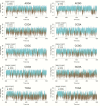
Twist distribution and correlation between twist and the possible states of the ζ angles. On the left, the observed distribution is depicted in black and normal components obtained with BIC in red (LT component), and in green (HT component) respectively. Correlation between the twist of the central CG step and the four states of the ζ angle at the 3′-junction are shown at the right of the distributions.
Figure 7.
Figure 7.
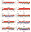
Time evolution of the twist at the CG step and the formation of the intra-molecular CH···O interaction. Results for the 10 possible tetranucleotides simulated in K+Cl.
Figure 8.
Figure 8.
Hydrogen bond AIM analysis for the TCGA tetranucleotide in the BII/BII conformation. The atoms labeled as C44, H45 and O30 represent the C8, H8 and O3′ atoms of the flanking purine. The bond critical points are evidenced by gray dots. The nuclear critical points (located at the position of the nuclei) are shown by black dots, while the basin paths and the gradient field are depicted by gray lines. The bond paths, defined by the chosen two-dimensional projection (plane), are shown by black lines.
Figure 9.
Figure 9.
PMF simulations performed with sodium on the ACGA tetranucleotide. All the possible ζ transitions from g-g- to tt that go through an intermediate state were considered.
Figure 10.
Figure 10.
Schematic timeline of the concerted events that start with the entering of the cations in the minor grooves of the CG steps. The events are measured before a ζ transition when going from g-/g- to t/t. The arrows in red correspond to events that occur in all the tetranucleotides. Yellow arrows correspond to events that only occur when a purine is flanking the CG step at the 3′ side.

References

    1. Franklin R.E., Gosling R.G. Molecular configuration in sodium thymonucleate. 1953. Nature. 2003;421:400–401. discussion 396. - PubMed
    1. Fuller W., Forsyth T., Mahendrasingam A. Water-DNA interactions as studied by X-ray and neutron fibre diffraction. Philos. Trans. R. Soc. Lond. B Biol. Sci. 2004;359:1237–1247. discussion 1247–1248. - PMC - PubMed
    1. Kypr J., Kejnovská I., Renciuk D., Vorlícková M. Circular dichroism and conformational polymorphism of DNA. Nucleic Acids Res. 2009;37:1713–1725. - PMC - PubMed
    1. Radhakrishnan I., Patel D.J. DNA triplexes: solution structures, hydration sites, energetics, interactions, and function. Biochemistry. 1994;33:11405–11416. - PubMed
    1. Bernués J., Azorín F. Triple-stranded DNA. In: Eckstein F., Lilley D.J., editors. Nucleic Acids and Molecular Biology SE - 1. Springer Berlin Heidelberg: Nucleic Acids and Molecular Biology; 1995. pp. 1–21.

Publication types