Comparative Study
. 2014 Nov 10;42(20):e155.
doi: 10.1093/nar/gku836. Epub 2014 Sep 15.

Comparison of TALE designer transcription factors and the CRISPR/dCas9 in regulation of gene expression by targeting enhancers

Affiliations
Comparative Study

Comparison of TALE designer transcription factors and the CRISPR/dCas9 in regulation of gene expression by targeting enhancers

Xuefei Gao et al. Nucleic Acids Res. .

Abstract

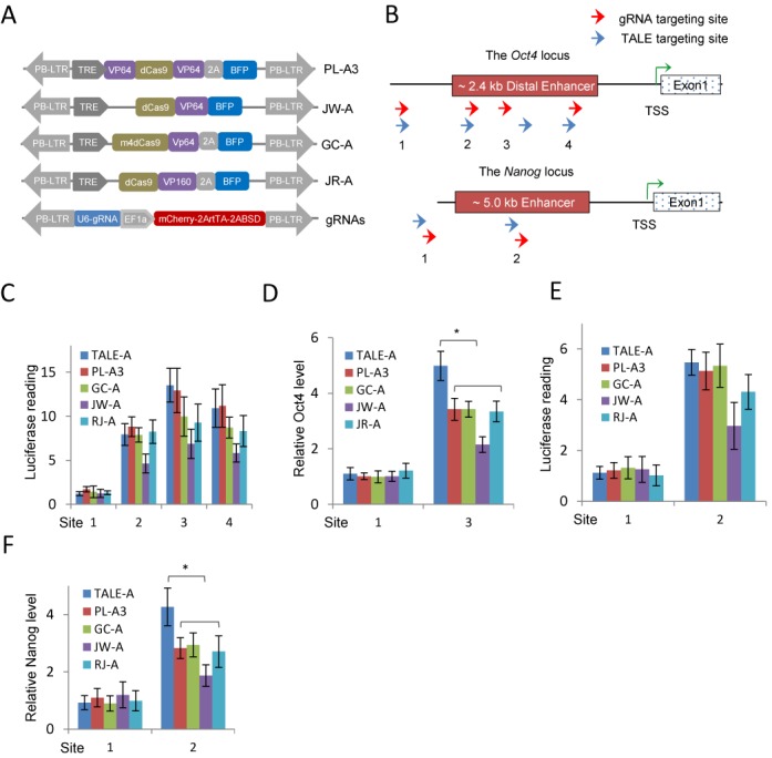
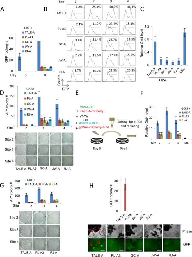
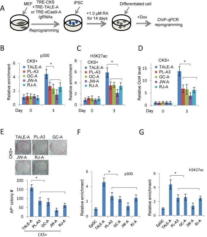
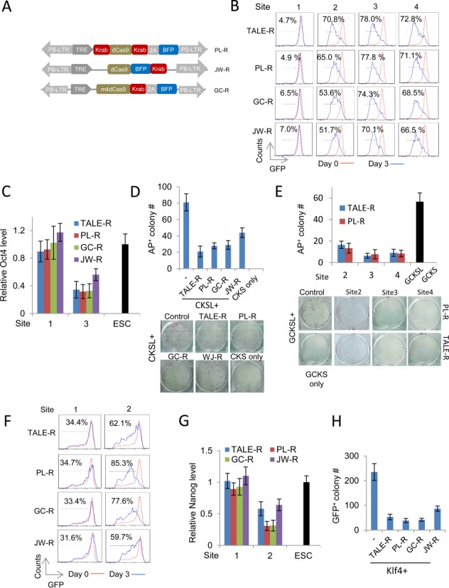
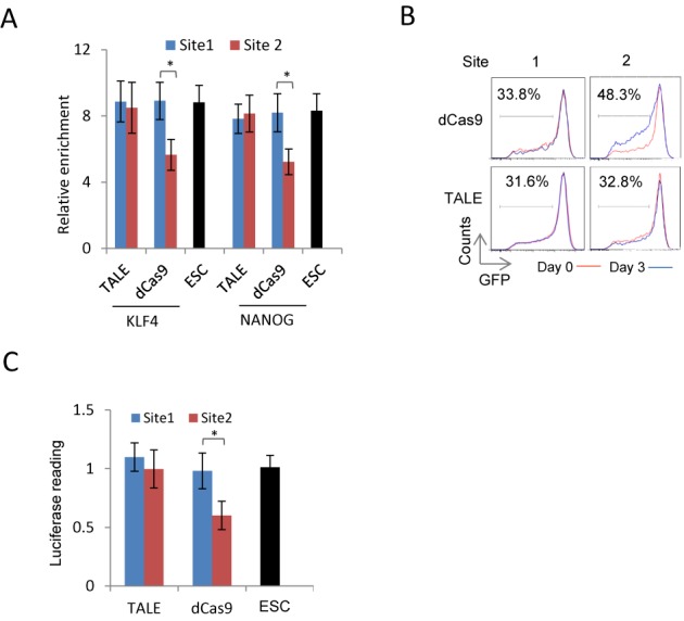
The transcription activator-like effectors (TALEs) and the RNA-guided clustered regularly interspaced short palindromic repeat (CRISPR) associated protein (Cas9) utlilize distinct molecular mechanisms in targeting site recognition. The two proteins can be modified to carry additional functional domains to regulate expression of genomic loci in mammalian cells. In this study, we have compared the two systems in activation and suppression of the Oct4 and Nanog loci by targeting their enhancers. Although both are able to efficiently activate the luciferase reporters, the CRISPR/dCas9 system is much less potent in activating the endogenous loci and in the application of reprogramming somatic cells to iPS cells. Nevertheless, repression by CRISPR/dCas9 is comparable to or even better than TALE repressors. We demonstrated that dCas9 protein binding results in significant physical interference to binding of native transcription factors at enhancer, less efficient active histone markers induction or recruitment of activating complexes in gene activation. This study thus highlighted the merits and drawbacks of transcription regulation by each system. A combined approach of TALEs and CRISPR/dCas9 should provide an optimized solution to regulate genomic loci and to study genetic elements such as enhancers in biological processes including somatic cell reprogramming and guided differentiation.


Figures

Figure 1.
Figure 1.
Activation of the mouse Oct4 and Nanog loci by TALE-As and dCas9-As/gRNAs. (A) Schematic diagram of the dCas9-As evaluated in this study. In all cases, blue florescent protein (BFP) was used to track the expression of dCas9-As. gRNA expression is controlled by U6 promoter, and EF1a-mCherry is used to detect the integration of the vector into the genome. A blasticidin resistance and the reverse tetracycline transactivator (rtTA) cassettes were also linked to the mCherry cassette by T2A peptides. PB-5TR and PB-3TR are the two terminal repeat sequences of the piggyBac (PB) transposon. (B) Schematic diagram of the TALE and dCas9/gRNA targeting sites at the mouse Oct4 and Nanog enhancers. Red arrows indicate the gRNA targeting sites and the blue arrows mark the TALE sites. (C) Activation of the Oct4 distal enhancer luciferase reporter by the TALE-As and dCas9-As/gRNAs. (D) qRT-PCR analysis of the Oct4 mRNA levels in MEFs expressing the TALE-As or dCas9-As/gRNAs. (E) Activation of the Nanog enhancer luciferase reporter by the TALE-As and dCas9-As/gRNAs. (F) qRT-PCR analysis of the Nanog mRNA levels in EpiSCs expressing the TALE-As or dCas9-As/gRNAs. All gene expression values were normalized to Gapdh. Results were representative of three independent experiments and were presented as ±SD, n = 3. *P < 0.05.
Figure 2.
Figure 2.
Somatic reprogramming to pluripotency by TALE-A or dCas9-As/gRNAs. (A) Comparison of GFP+ colony number reprogrammed from Oct4-GFP MEFs by CKS factors plus TALE-A or dCas9-As targeting at Site 3 of the Oct4 distal enhancer on day 5 and 8 post transfection. (B) Flow cytometry analysis of GFP+ cells in Oct4-GFP MEFs on day 8 by CKS factors plus TALE-As or dCas9-As targeting at Sites 1–4 of the Oct4 distal enhancer. (C) The endogenous Oct4 mRNA expression levels in GFP+Oct4-GFP MEFs expressing CKS factors plus TALE-A or dCas9-As targeting at Site 3. (D) The number of AP+ colonies reprogrammed from Oct4-GFP MEFs as in (B). (E) Schematic diagram showing the design of reprogramming experiment with FACS-sorted wild-type MEFs to control for the technical variability in primary transfection. Green: GFP-tagged CKS vector, blue: BFP-tagged dCas9-As vector and red: mCherry-tagged TALE-As or gRNAs-rtTA vector. (F) The endogenous Oct4 expression levels in the FACS-sorted wild-type MEFs expressing the TALE-A or dCas9-As targeting at Site 2, 3 and 4 of the Oct4 distal enhancer. (G) The number of AP+ colony reprogrammed from the FACS-sorted wild-type MEFs (20 000 cells/well) as in (F). (H) Reprogramming Oct4-GFP reporter EpiSCs by TALE-As and dCas9-As/gRNAs targeting at the Site 2 of the Nanog enhancer. The iPSC colonies were indicated by red arrows. The number of GFP+ surviving colony was quantified 14 days after 2i media selection. Scale bars: 200.0 μm. Results were representative of three independent experiments and were presented as ±SD, n = 3.
Figure 3.
Figure 3.
Epigenetic changes induced by TALE-As and dCas9-As/gRNAs in secondary MEF (AE) and EpiSCs (F and G) reprogramming experiments. (A) Schematic diagram showing the experimental design of the secondary MEF reprogramming experiment. (B and C) ChIP-qPCR analysis of p300 and H3K27Ac enrichment at the Oct4 distal enhancer. (D) The endogenous Oct4 mRNA levels in RA-differentiated cells after 3 days of Dox induction. The relative enrichments were normalized to IgG, and a genomic region at the Tyr locus was used as a control region. (E) Quantification of secondary MEF reprogramming efficiency by TALE-As and dCas9-As/gRNAs. 1500 differentiated cells were plated into each well of a 6-well plate and AP+ colonies were scored 12 days after Dox induction. (F and G) Relative p300 and H3K27Ac enrichment at the Nanog enhancer in Oct4-GFP EpiSCs expressing TALE-A or dCas9-As targeting at the Site 2 of the Nanog 5 kb enhancer detected by ChIP-qPCR detection. Results were representative of three independent experiments and were presented as ±SD, n = 3. *P < 0.05.
Figure 4.
Figure 4.
Repression of the Oct4 and Nanog loci by TALE-Rs and dCas9-Rs/gRNAs. (A) Schematic diagram of the dCas9-R designs evaluated in this study. In all cases, blue florescent protein (BFP) was used to track the expression of dCas9-R. The gRNA vector used was the same as described in Figure 1. (B) Repression of the endogenous Oct4 locus in Oct4-GFP ES cells indicated by the reduction of GFP intensity in flow cytometric analysis on day 0 and day 3 of expression of the TALE-Rs or dCas9-Rs/gRNAs targeting at the Sites 1–4 of the Oct4 distal enhancer (Gated mCherry+ for TALEs and mCherry+/BFP+ for dCas9-As/gRNAs). (C) Comparison of Oct4 expression levels in Oct4-GFP ES cells expressing the TALE-Rs or dCas9-Rs targeting at the Site 1 or Site 3 of the Oct4 distal enhancer by qRT-PCR. (D) The repressive effect of TALE-R and dCas9-Rs/gRNAs targeting at the Site 3 of the Oct4 distal enhancer on MEF reprogramming. MEFs were reprogrammed with Dox inducible CKS and Lrh1 (CKSL) factors together with the TALE-R or dCas9-R/gRNAs as in (C). ‘CKSL+’ indicates that all transfections have CKSL. ‘−’ is the CKSL only control (no repressor). ‘CKS only’ is the reprogramming negative control. (E) Reprogramming using FACS-sorted MEFs (as described in 2E) to control transfection variability. Wild-type MEFs were transfected and sorted on day 2 for GFP+/mCherry+ in the TALE-R transfection and for GFP+/mCherry+/BFP+ in dCas9-R/gRNAs (PL-R) experiments. Both TALE-Rs and dCas9-R/gRNAs (PL-R) targeted the Site 2–4 of the Oct4 distal enhancer. Sorted MEFs were re-plated (20 000 cells/ well) for reprogramming and iPSC colonies were scored by AP staining. (F) The repressive effect of TALE-Rs or dCas9-Rs/gRNAs targeting at the Sites 1–2 of the Nanog 5 kb enhancer in Nanog-GFP ES cells. Site 1 is located outside the enhancer region whereas Site 2 is inside. The Nanog repression was demonstrated by the increase of the GFPlow/dim fraction in Nanog-GFP ES cells. (G) Endogenous Nanog mRNA levels in Nanog-GFP ES cells expressing the TALE-Rs or dCas9-Rs targeting at the Sites 1–2. (H) Repression of Klf4-mediated EpiSC reprogramming to iPSCs by the TALE-R and dCas9-Rs targeting at the Site 2 of the Nanog 5 kb enhancer. ‘Klf4+’ refers to the transfections combined with Klf4 and ‘−’ is the Klf4 control (no effector). Results were representative of three independent experiments and were presented as ±SD, n = 3.
Figure 5.
Figure 5.
Interference of dCas9 protein on native transcription factor binding at the Nanog enhancer. (A) Change of KLF4 and NANOG binding at the 5 kb Nanog enhancer region induced by regulatory-domain-free dCas9 or TALE protein targeting at the Sites 1–2 detected by ChIP-qPCR in mouse ES cells. WT: ES cells transfected with an empty control vector. (B) Change of the GFPlow/dim fraction in Nanog-GFP ES cells induced by regulatory-domain-free dCas9 and TALE protein targeting at the Site 1–2 of the Nanog enhancer on days 0 and 3 after transfection. (C) Nanog enhancer luciferase reporter activities in mouse ES cells expressing regulatory-domain-free dCas9 or TALE protein targeting at the Sites 1–2. Results were representative of three independent experiments and were presented as ±SD, n = 3. *P < 0.05.

References

    1. Takahashi K., Yamanaka S. Induction of pluripotent stem cells from mouse embryonic and adult fibroblast cultures by defined factors. Cell. 2006;126:663–676. - PubMed
    1. Theunissen T.W., van Oosten A.L., Castelo-Branco G., Hall J., Smith A., Silva J.C. Nanog overcomes reprogramming barriers and induces pluripotency in minimal conditions. Curr. Biol. 2011;21:65–71. - PMC - PubMed
    1. Guo G., Yang J., Nichols J., Hall J.S., Eyres I., Mansfield W., Smith A. Klf4 reverts developmentally programmed restriction of ground state pluripotency. Development. 2009;136:1063–1069. - PMC - PubMed
    1. Brons I.G., Smithers L.E., Trotter M.W., Rugg-Gunn P., Sun B., Chuva de Sousa Lopes S.M., Howlett S.K., Clarkson A., Ahrlund-Richter L., Pedersen R.A., et al. Derivation of pluripotent epiblast stem cells from mammalian embryos. Nature. 2007;448:191–195. - PubMed
    1. Li R., Liang J., Ni S., Zhou T., Qing X., Li H., He W., Chen J., Li F., Zhuang Q., et al. A mesenchymal-to-epithelial transition initiates and is required for the nuclear reprogramming of mouse fibroblasts. Cell Stem Cell. 2010;7:51–63. - PubMed

Publication types

Substances