. 2014 Dec:77:21-9.
doi: 10.1016/j.freeradbiomed.2014.08.025. Epub 2014 Sep 16.

A time course of NADPH-oxidase up-regulation and endothelial nitric oxide synthase activation in the hippocampus following neurotrauma

Affiliations

A time course of NADPH-oxidase up-regulation and endothelial nitric oxide synthase activation in the hippocampus following neurotrauma

Mubeen A Ansari et al. Free Radic Biol Med. 2014 Dec.

Abstract

Nicotinamide adenine dinucleotide phosphate oxidase (NADPH-oxidase; NOX) is a complex enzyme responsible for increased levels of reactive oxygen species (ROS), superoxide (O2(•-)). NOX-derived O2(•-) is a key player in oxidative stress and inflammation-mediated multiple secondary injury cascades (SIC) following traumatic brain injury (TBI). The O2(•-) reacts with nitric oxide (NO), produces various reactive nitrogen species (RNS), and contributes to apoptotic cell death. Following a unilateral cortical contusion, young adult rats were killed at various times postinjury (1, 3, 6, 12, 24, 48, 72, and 96 h). Fresh tissue from the hippocampus was analyzed for NOX activity, and level of O2(•-). In addition we evaluated the translocation of cytosolic NOX proteins (p67(Phox), p47(Phox), and p40(Phox)) to the membrane, along with total NO and the activation (phosphorylation) of endothelial nitric oxide synthase (p-eNOS). Results show that both enzymes and levels of O2(•-) and NO have time-dependent injury effects in the hippocampus. Translocation of cytosolic NOX proteins into membrane, NOX activity, and O2(•-) were also increased in a time-dependent fashion. Both NOX activity and O2(•-) were increased at 6 h. Levels of p-eNOS increased within 1h, with significant elevation of NO at 12h post-TBI. Levels of NO failed to show a significant association with p-eNOS, but did associate with O2(•-). NOX up-regulation strongly associated with both the levels of O2(•-) and the total NO. The initial 12 h post-TBI are very important as a possible window of opportunity to interrupt SIC. It may be important to selectively target the translocation of cytosolic subunits for the modulation of NOX function.

Keywords: Free radicals; NADPH-oxidase; Secondary injury cascades; Traumatic brain injury.


Figures

Figure 1
Figure 1
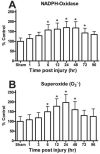
Time course for the activity of NADPH Oxidase (NOX) and levels of superoxide (O2.−) in the ipsilateral hippocampus following a moderate cortical contusion. (A) A significant elevation in the NOX activity occurred at 6 h after injury, with the maximum NOX activity observed at 24 h. Levels of NOX derived O2.− also demonstrated a time-dependent increase with significant changes within 6 h post trauma and maximum increases at 24 h post injury. (B) Each bar represents the group mean ± SD of six animals/group. *p < 0.05 versus sham operates.
Figure 2
Figure 2
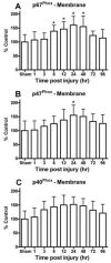
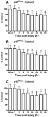
Key cytosolic NOX subunit proteins were quantitatively assessed in the hippocampus with immunobloting followed by Western-blot. (A) The levels of cytosolic p67Phox, p47Phox, and p40Phox were increased in membrane fractions and (B) declined in cytosolic fraction, demonstrating a substantial translocation of these subunits. Na+/K+-ATPase and GAPDH show equal protein used for membrane and cytosolic Western-blots. Time dependent changes in different NOX proteins in ipsilateral hippocampus membrane fraction following a moderate cortical contusion. Cytosolic NOX protein, p67Phox (C), was significantly increased early (6 h) in the membrane fraction of the hippocampus following TBI. The maximum increase of p67Phox in the membrane fraction was at 24 h and continued at 96 h post TBI. In a similar fashion, other key cytosolic NOX protein, p47Phox (E), and p40Phox (G) were also increased in membrane fraction of ipsilateral hippocampus as compared sham animals. Each bar represents the group mean ± SD of six animals/group. *p < 0.05 versus sham operates. Levels of NOX proteins, p67Phox (D), p47Phox (F), and p40Phox (H) significantly decreased in cytosolic fraction of the hippocampus following TBI. Result indicates there was a time dependently sifting/translocation of cytosolic component to the membrane for the up-regulation of NOX and increase production of O2 after TBI. Each bar represents the group mean ± SD of six animals/group. *p < 0.05 versus sham operated rats.
Figure 3
Figure 3
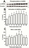
Time-dependent changes in hippocampal phosphorylated eNOS (p-eNOS) and level total nitric oxide (NO) after a moderate cortical contusion. (A) Western-blot analysis shows p-eNOS was significant increase in the ipsilateral hippocampus, as early as 1 h and maintained elevated levels at 96 h post trauma (B). Total NO levels in ipsilateral hippocampus were significantly increased at 24 h after TBI (C). Each bar represents the group mean ± SD of six animals/group. *p < 0.05 versus sham operated rats.
Figure 4
Figure 4
Scatterplot (A, C, E) showing the relationship between key NOX subunits (p-67Phox, p-47Phox, & p-40Phox) in the membrane fraction and changes in NOX activity Increases in the subunits significantly correlated with the elevation in total NOX activity. Scatterplot (B, D, F) shows the relationship between key NOX subunits (p-67Phox, p-47Phox, & p-40Phox) in the membrane fraction and changes in O2.− activity. Lines are shown to represent the direction of the correlation.
Figure 5
Figure 5
Scatterplot showing the significant relationship between levels of NOX activity and O2.− in the ipsilateral hippocampus following a moderate TBI (A). As the NOX levels increased they were paralleled by an increase in O2.−. NOX levels also significantly correlated with changes in the levels of NO (C). Levels of NO in the hippocampus following the moderate TBI significantly associated with increased O2.−. The analysis failed to demonstrate a significant correlation between p-eNOS levels and NO following the injury (B). Lines are shown to represent the direction of the correlation.
Figure 6
Figure 6
Figure 7
Figure 7

Similar articles

Cited by

References

    1. Langlois JA, Marr A, Mitchko J, Johnson RL. Tracking the silent epidemic and educating the public: CDC's traumatic brain injury-associated activities under the TBI Act of 1996 and the Children's Health Act of 2000. J Head Trauma Rehabil. 2005;20:196–204. - PubMed
    1. Bayir H, Kagan VE, Clark RS, Janesko-Feldman K, Rafikov R, Huang Z, Zhang X, Vagni V, Billiar TR, Kochanek PM. Neuronal NOS-mediated nitration and inactivation of manganese superoxide dismutase in brain after experimental and human brain injury. J Neurochem. 2007;101:168–181. - PubMed
    1. Hall ED, Detloff MR, Johnson K, Kupina NC. Peroxynitrite-mediated protein nitration and lipid peroxidation in a mouse model of traumatic brain injury. J Neurotrauma. 2004;21:9–20. - PubMed
    1. Siesjo BK. Calcium-mediated processes in neuronal degeneration. Ann N Y Acad Sci. 1994;747:140–161. - PubMed
    1. Gilmer LK, Ansari MA, Roberts KN, Scheff SW. Age-related mitochondrial changes after traumatic brain injury. J Neurotrauma. 2010;27:939–950. - PMC - PubMed

Publication types

LinkOut - more resources