

Left-ventricular mechanical activation and aortic-arch orientation recovered from magneto-hydrodynamic voltages observed in 12-lead ECGs obtained inside MRIs: a feasibility study
- PMID: 25224074
- PMCID: PMC4241154
- DOI: 10.1007/s10439-014-1109-2
Left-ventricular mechanical activation and aortic-arch orientation recovered from magneto-hydrodynamic voltages observed in 12-lead ECGs obtained inside MRIs: a feasibility study
Abstract
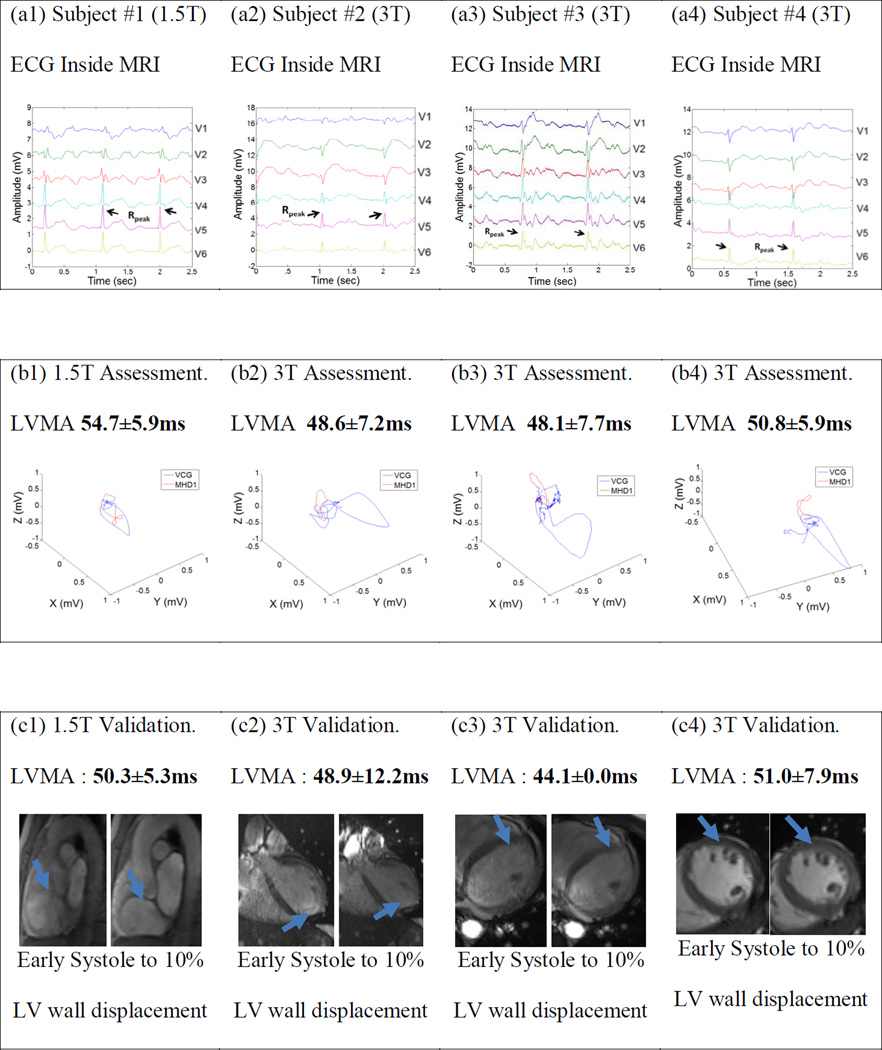
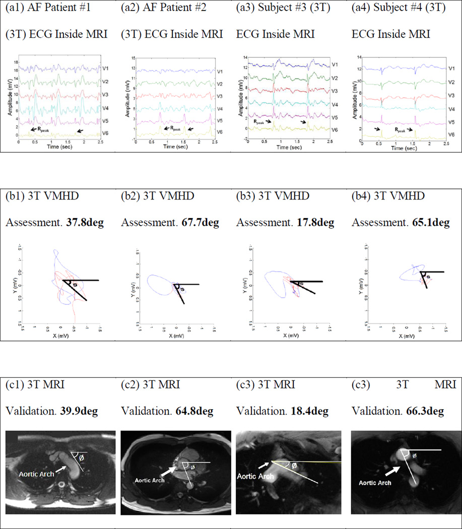
To explore use of the Magnetohydrodynamic Voltage (VMHD), observed in intra-MRI 12-lead electrocardiograms (ECG), to indicate the timing of the onset of left-ventricular mechanical activation (LVMA) and the orientation of the aortic-arch (AAO). Blood flow through the aortic arch during systole, in the presence of the MRI magnetic field (B 0), generates VMHD. Since the magnitude and direction of VMHD are determined by the timing and directionality of blood flow relative to B 0, we hypothesized that clinically useful measures, LVMA and AAO, could be extracted from temporal and vectorial VMHD characteristics. VMHD signals were extracted from 12-lead ECG traces by comparing traces obtained inside and outside the MRI scanner. VMHD was converted into the Vectorcardiogram frame of reference. LVMA was quantified in 1 subject at 1.5T and 3 subjects at 3T, and the result compared to CINE MRI. AAO was inferred for 4 subjects at 3T and compared to anatomical imaging of the aortic arch orientation in the transverse plane. A < 10% error was observed in LVMA measurements, while a < 3° error was observed in aortic arch orientation measurements. The temporal and vectorial nature of VMHD is useful in estimating these clinically relevant parameters.
Figures


References
-
- Gupta A, Weeks AR, Richie SM. Simulation of elevated T-waves of an ECG inside a static magnetic field (MRI) IEEE transactions on bio-medical engineering. 2008;55:1890–1896. - PubMed
-
- Blandford R, Thorne K. Applications of Classical Physics. CA: CalTech; 2004. Magnetohydrodynamics.
-
- Krug J, Rose G. Magnetohydrodynamic distortions of the ECG in different MR scanner configurations. Computing in Cardiology. 2011:769–772.
-
- Birkholz T, Schmid M, Nimsky C, Schuttler J, Schmitz B. ECG artifacts during intraoperative high-field MRI scanning. Journal of neurosurgical anesthesiology. 2004;16:271–276. - PubMed
-
- Nijm G, Swiryn S, Larson A, Sahakian A. Characterization of the magnetohydrodynamic effect as a signal from the surface electrocardiogram during cardiac magnetic resonance imaging. IEEE; 2006.
Publication types
MeSH terms
Grants and funding
LinkOut - more resources
Full Text Sources
Other Literature Sources
