

Tumor-suppressive miR148a is silenced by CpG island hypermethylation in IDH1-mutant gliomas
- PMID: 25224277
- PMCID: PMC4233178
- DOI: 10.1158/1078-0432.CCR-14-0234
Tumor-suppressive miR148a is silenced by CpG island hypermethylation in IDH1-mutant gliomas
Abstract
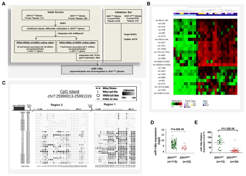
Purpose: IDH1/2-mutant gliomas harbor a distinct glioma-CpG island methylation phenotype (G-CIMP) that may promote the initiation and progression of secondary pathway gliomas by silencing tumor-suppressive genes. The potential role of tumor-suppressive microRNAs (miRNA; miR) in this process is not understood.
Experimental design: To identify potential tumor-suppressive miRNA hypermethylated in glioma, the methylation profiles of IDH1/2(WT) gliomas (n = 11) and IDH1(MUT) glioma (n = 20) were compared by using massively parallel reduced representation bisulfite sequencing (RRBS). The methylation status of selected miRNA was validated by using targeted bisulfite sequencing (BiSEQ) in a large cohort of glioma tissue samples including 219 IDH1(WT) and 72 IDH1/2(MUT) samples. The expression of selected miRNAs was determined by using the TaqMan qPCR. Functional analyses of miR148a were conducted and target genes were identified.
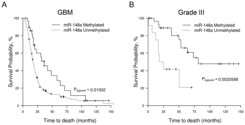
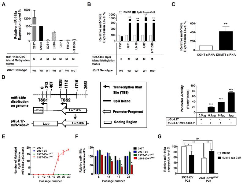
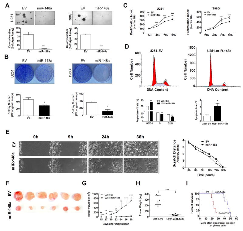
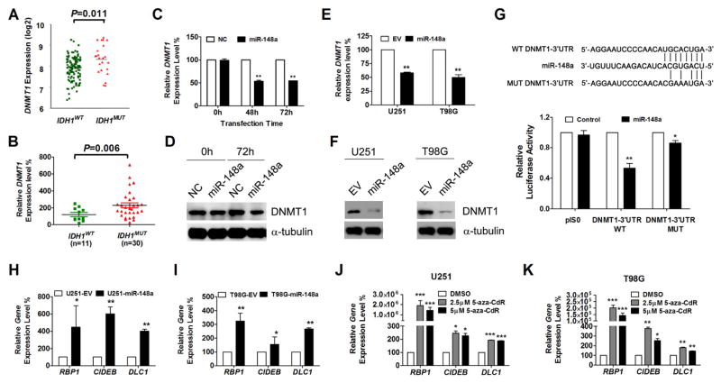
Results: We identify miR148a as a novel, G-CIMP-associated miRNA whose methylation is tightly correlated with IDH1 mutation and associated with improved survival in patients with malignant glioma. We confirm that downregulation of miR148a can occur via DNA methylation. We demonstrate that IDH1 mutation provides a mechanism of miR148a methylation and downregulation, and that restoration of miR148a reduced tumorigenic properties of glioma cells, possibly by targeting DNMT1.
Conclusions: We identify miR148a as a novel G-CIMP-associated miRNA, and provide results suggesting that miR148a restoration may have therapeutic implications.
©2014 American Association for Cancer Research.
Conflict of interest statement
Figures
Similar articles
-
Identification of retinol binding protein 1 promoter hypermethylation in isocitrate dehydrogenase 1 and 2 mutant gliomas.J Natl Cancer Inst. 2012 Oct 3;104(19):1458-69. doi: 10.1093/jnci/djs357. Epub 2012 Sep 3. J Natl Cancer Inst. 2012. PMID: 22945948 Free PMC article.
-
SPINT2 is hypermethylated in both IDH1 mutated and wild-type glioblastomas, and exerts tumor suppression via reduction of c-Met activation.J Neurooncol. 2019 May;142(3):423-434. doi: 10.1007/s11060-019-03126-x. Epub 2019 Mar 5. J Neurooncol. 2019. PMID: 30838489 Free PMC article.
-
CRISPR Editing of Mutant IDH1 R132H Induces a CpG Methylation-Low State in Patient-Derived Glioma Models of G-CIMP.Mol Cancer Res. 2019 Oct;17(10):2042-2050. doi: 10.1158/1541-7786.MCR-19-0309. Epub 2019 Jul 10. Mol Cancer Res. 2019. PMID: 31292202 Free PMC article.
-
The Cancer Genome Atlas expression profiles of low-grade gliomas.Neurosurg Focus. 2014 Apr;36(4):E23. doi: 10.3171/2012.12.focus12351. Neurosurg Focus. 2014. PMID: 24812719 Review.
-
Overview of DNA methylation in adult diffuse gliomas.Brain Tumor Pathol. 2019 Apr;36(2):84-91. doi: 10.1007/s10014-019-00339-w. Epub 2019 Apr 1. Brain Tumor Pathol. 2019. PMID: 30937703 Review.
Cited by
-
Partial erosion on under-methylated regions and chromatin reprogramming contribute to oncogene activation in IDH mutant gliomas.Epigenetics Chromatin. 2023 Apr 28;16(1):13. doi: 10.1186/s13072-023-00490-x. Epigenetics Chromatin. 2023. PMID: 37118755 Free PMC article.
-
New developments in the pathogenesis and therapeutic targeting of the IDH1 mutation in glioma.Int J Med Sci. 2015 Jan 20;12(3):201-13. doi: 10.7150/ijms.11047. eCollection 2015. Int J Med Sci. 2015. PMID: 25678837 Free PMC article. Review.
-
IGFBP2 expression predicts IDH-mutant glioma patient survival.Oncotarget. 2017 Jan 3;8(1):191-202. doi: 10.18632/oncotarget.13329. Oncotarget. 2017. PMID: 27852048 Free PMC article.
-
MiR-148a increases glioma cell migration and invasion by downregulating GADD45A in human gliomas with IDH1 R132H mutations.Oncotarget. 2017 Apr 11;8(15):25345-25361. doi: 10.18632/oncotarget.15867. Oncotarget. 2017. PMID: 28445981 Free PMC article.
-
Commercial rodent diets differentially regulate autoimmune glomerulonephritis, epigenetics and microbiota in MRL/lpr mice.Int Immunol. 2017 Jun 1;29(6):263-276. doi: 10.1093/intimm/dxx033. Int Immunol. 2017. PMID: 28637300 Free PMC article.
References
-
- Behin A, Hoang-Xuan K, Carpentier AF, Delattre JY. Primary brain tumours in adults. Lancet. 2003;361:323–31. - PubMed
-
- Ohgaki H, Kleihues P. The definition of primary and secondary glioblastoma. Clinical cancer research : an official journal of the American Association for Cancer Research. 2013;19:764–72. - PubMed
Publication types
MeSH terms
Substances
Grants and funding
LinkOut - more resources
Full Text Sources
Other Literature Sources
Medical
Research Materials
Miscellaneous
