Human T-follicular helper and T-follicular regulatory cell maintenance is independent of germinal centers
- PMID: 25224411
- PMCID: PMC4208282
- DOI: 10.1182/blood-2014-07-585976
Human T-follicular helper and T-follicular regulatory cell maintenance is independent of germinal centers
Abstract
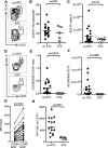
The monoclonal anti-CD20 antibody rituximab (RTX) depletes B cells in the treatment of lymphoma and autoimmune disease, and contributes to alloantibody reduction in transplantation across immunologic barriers. The effects of RTX on T cells are less well described. T-follicular helper (Tfh) cells provide growth and differentiation signals to germinal center (GC) B cells to support antibody production, and suppressive T-follicular regulatory (Tfr) cells regulate this response. In mice, both Tfh and Tfr are absolutely dependent on B cells for their formation and on the GC for their maintenance. In this study, we demonstrate that RTX treatment results in a lack of GC B cells in human lymph nodes without affecting the Tfh or Tfr cell populations. These data demonstrate that human Tfh and Tfr do not require an ongoing GC response for their maintenance. The persistence of Tfh and Tfr following RTX treatment may permit rapid reconstitution of the pathological GC response once the B-cell pool begins to recover. Strategies for maintaining remission after RTX therapy will need to take this persistence of Tfh into account.
© 2014 by The American Society of Hematology.
Figures


Similar articles
-
Interleukin-21 promotes germinal center reaction by skewing the follicular regulatory T cell to follicular helper T cell balance in autoimmune BXD2 mice.Arthritis Rheumatol. 2014 Sep;66(9):2601-12. doi: 10.1002/art.38735. Arthritis Rheumatol. 2014. PMID: 24909430 Free PMC article.
-
Decreased T Follicular Regulatory Cell/T Follicular Helper Cell (TFH) in Simian Immunodeficiency Virus-Infected Rhesus Macaques May Contribute to Accumulation of TFH in Chronic Infection.J Immunol. 2015 Oct 1;195(7):3237-47. doi: 10.4049/jimmunol.1402701. Epub 2015 Aug 21. J Immunol. 2015. PMID: 26297764 Free PMC article.
-
A distinct subpopulation of CD25- T-follicular regulatory cells localizes in the germinal centers.Proc Natl Acad Sci U S A. 2017 Aug 1;114(31):E6400-E6409. doi: 10.1073/pnas.1705551114. Epub 2017 Jul 11. Proc Natl Acad Sci U S A. 2017. PMID: 28698369 Free PMC article.
-
T cell immune response within B-cell follicles.Adv Immunol. 2019;144:155-171. doi: 10.1016/bs.ai.2019.08.008. Epub 2019 Sep 16. Adv Immunol. 2019. PMID: 31699216 Review.
-
T Follicular Regulatory Cells and Antibody Responses in Transplantation.Transplantation. 2018 Oct;102(10):1614-1623. doi: 10.1097/TP.0000000000002224. Transplantation. 2018. PMID: 29757907 Free PMC article. Review.
Cited by
-
Effector and regulatory B cells in immune-mediated kidney disease.Nat Rev Nephrol. 2019 Jan;15(1):11-26. doi: 10.1038/s41581-018-0074-7. Nat Rev Nephrol. 2019. PMID: 30443016 Review.
-
T follicular regulatory cells infiltrate the human airways during the onset of acute respiratory distress syndrome and regulate the development of B regulatory cells.Immunol Res. 2018 Aug;66(4):548-554. doi: 10.1007/s12026-018-9014-7. Immunol Res. 2018. PMID: 30051220 Free PMC article.
-
T Follicular Helper Cells in Autoimmune Disorders.Front Immunol. 2018 Jul 17;9:1637. doi: 10.3389/fimmu.2018.01637. eCollection 2018. Front Immunol. 2018. PMID: 30065726 Free PMC article. Review.
-
MicroRNA signatures and Foxp3+ cell count correlate with relapse occurrence in follicular lymphoma.Oncotarget. 2018 Apr 13;9(28):19961-19979. doi: 10.18632/oncotarget.24987. eCollection 2018 Apr 13. Oncotarget. 2018. PMID: 29731996 Free PMC article.
-
PDL1 blockage increases fetal resorption and Tfr cells but does not affect Tfh/Tfr ratio and B-cell maturation during allogeneic pregnancy.Cell Death Dis. 2020 Feb 12;11(2):119. doi: 10.1038/s41419-020-2313-7. Cell Death Dis. 2020. PMID: 32051396 Free PMC article.
References
-
- MacLennan IC. Germinal centers. Annu Rev Immunol. 1994;12:117–139. - PubMed
-
- Meyer-Hermann M, Maini P, Iber D. An analysis of B cell selection mechanisms in germinal centers. Math Med Biol. 2006;23(3):255–277. - PubMed
-
- Victora GD, Nussenzweig MC. Germinal centers. Annu Rev Immunol. 2012;30:429–457. - PubMed
-
- Vinuesa CG, Cyster JG. How T cells earn the follicular rite of passage. Immunity. 2011;35(5):671–680. - PubMed
Publication types
MeSH terms
Substances
Grants and funding
LinkOut - more resources
Full Text Sources
Other Literature Sources
Miscellaneous

