. 2014 Oct 30;124(18):2881-91.
doi: 10.1182/blood-2014-04-570440. Epub 2014 Sep 15.

α-1-Antitrypsin (AAT)-modified donor cells suppress GVHD but enhance the GVL effect: a role for mitochondrial bioenergetics

Affiliations

α-1-Antitrypsin (AAT)-modified donor cells suppress GVHD but enhance the GVL effect: a role for mitochondrial bioenergetics

A Mario Marcondes et al. Blood. .

Abstract

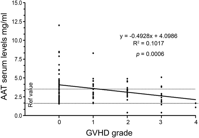
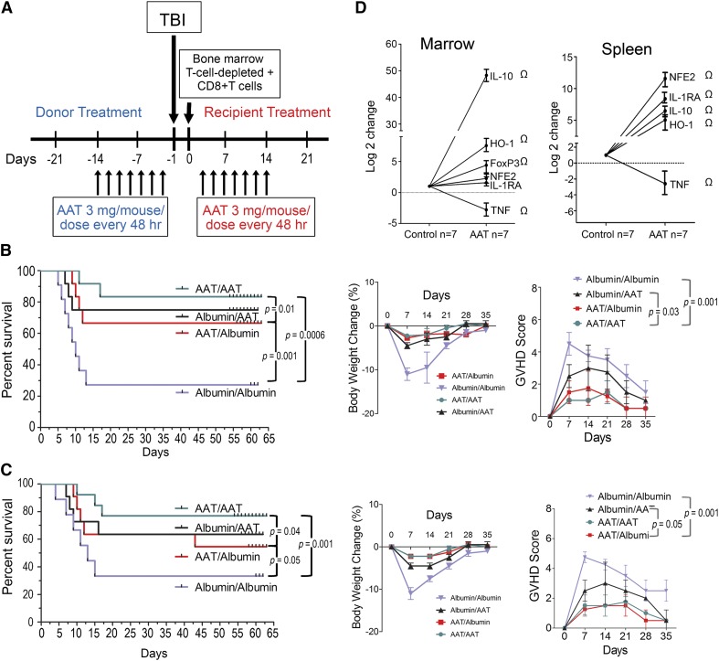
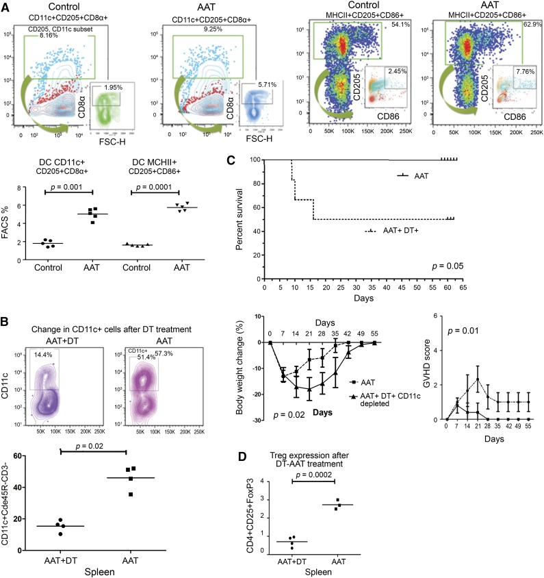
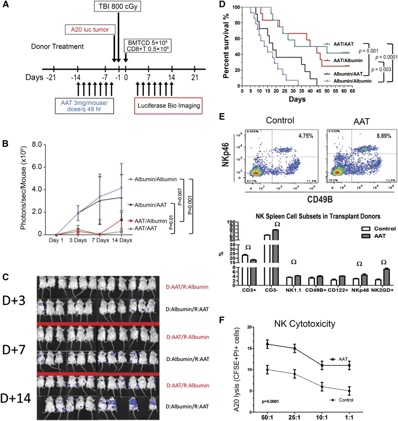
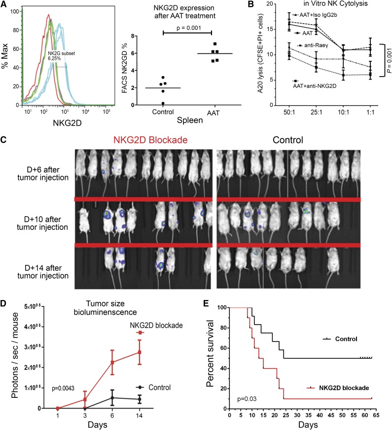
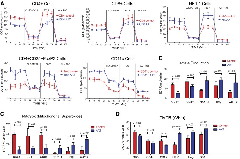
Hematopoietic cell transplantation is curative in many patients. However, graft-versus-host disease (GVHD), triggered by alloreactive donor cells, has remained a major complication. Here, we show an inverse correlation between plasma α-1-antitrypsin (AAT) levels in human donors and the development of acute GVHD in the recipients (n = 111; P = .0006). In murine models, treatment of transplant donors with human AAT resulted in an increase in interleukin-10 messenger RNA and CD8(+)CD11c(+)CD205(+) major histocompatibility complex class II(+) dendritic cells (DCs), and the prevention or attenuation of acute GVHD in the recipients. Ablation of DCs (in AAT-treated CD11c-DTR donors) decreased CD4(+)CD25(+)FoxP3(+) regulatory T cells to one-third and abrogated the anti-GVHD effect. The graft-versus-leukemia (GVL) effect of donor cells (against A20 tumor cells) was maintained or even enhanced with AAT treatment of the donor, mediated by an expanded population of NK1.1(+), CD49B(+), CD122(+), CD335(+) NKG2D-expressing natural killer (NK) cells. Blockade of NKG2D significantly suppressed the GVL effect. Metabolic analysis showed a high glycolysis-high oxidative phosphorylation profile for NK1.1(+) cells, CD4(+)CD25(+)FoxP3(+) T cells, and CD11c(+) DCs but not for effector T cells, suggesting a cell type-specific effect of AAT. Thus, via altered metabolism, AAT exerts effective GVHD protection while enhancing GVL effects.


Figures

Figure 1
Figure 1
Donor AAT levels and acute GVHD (n = 111) by day 100. Plasma AAT levels in human allogeneic transplant donors. Donor AAT levels and the development of GVHD (by grade) in the respective recipients by day 100. GVHD severity increased with decreasing AAT levels. Modeling GVHD severity as a linear variable, a trend test showed decreasing average AAT plasma levels with increasing GVHD scores (P = .0006). This same conclusion held for GVHD scores by day 20 (P = .01) and by day 30 (P = .002) (not shown).
Figure 2
Figure 2
Effects of AAT treatment of donor, recipient, or both on GVHD and posttransplant outcome. (A) Treatment scheme for both donor (blue) and recipient (red). (B) Results with C3H.SW (H-2b) donors and C57BL/6 (H 2bc) recipients (minor mismatch) and (C) results with C57BL/6 (H-2bKb) donors, and Balb/c (H-2d Kd) recipients (major mismatch). Either donor (AAT/albumin) or recipient (albumin/AAT), both (AAT/AAT) or neither (albumin/albumin) were treated with AAT; albumin served as control. Left panels show survival; middle and right panels show body weight changes and GVHD scores (n = 12 for each transplant condition). (D) Changes in cytokine levels in donor marrow and spleen following AAT treatment relative to albumin controls (determined by RT-PCR; n = 7 mice per group). Results are expressed as mean ± SEM (log2) from pooled tissue extracts (marrow and spleen preparations). Ω indicates P values of at least = .01 when compared with albumin-treated control cells.
Figure 3
Figure 3
Effect of AAT on donor cell subpopulations. (A) Increase in the proportion of DCs in spleens of AAT-treated (AAT) and albumin-treated (controls) donors. Shown are spleen cells labeled for CD11c+CD205+CD8+ (left panels), and for MHC class II+CD205+CD86+ (right panels); data on all mice are summarized in the graph. (B) Treatment of CD11c-DTR mice expressing the DT receptor with DT or PBS (control). Bone marrow and spleen cells were collected and stained for CD11c. Following 2 injections of DT, CD11c-expressing cells were 80% to 90% depleted (see “Materials and methods”). (C) Survival of C3H.SW (H 2bc) recipients of marrow plus spleen cells from C57BL/6 [H-2b] donors that had been treated with AAT (AAT) only or treated with AAT and also injected with DT. Recipients of DC-depleted donor cells had greater weight loss and higher GVHD scores (lower panels) than mice transplanted from donors not injected with DT (n = 7, each arm). (D) Treatment of donor mice transgenic for CD11c DTR with DT also significantly reduced the proportion of CD4+CD25+FoxP3+ Tregs.
Figure 4
Figure 4
Effect of AAT on NK cells and antitumor activity. (A) Treatment scheme: injection of A20 tumor cells (A20 luc tumor) and transplantation; C57BL/6 (H-2bKb) donors, Balb/c (H-2d Kd) recipients (major mismatch). (B) Either donors (D) or recipients (R) or both were treated with AAT (controls received albumin), resulting in four donor/recipient combinations: transplantation of cells from AAT-treated donors into recipients treated with albumin (AAT/albumin; red); transplantation of cells from albumin-treated donors into AAT-treated recipients (albumin/AAT; black); transplantation of cells from AAT-treated donors into AAT-treated recipients (AAT/AAT; green), and transplantation of cells from albumin-treated donors into albumin-treated recipients (albumin/albumin; purple). Photon reading of luciferase activity; higher readings indicate greater tumor volume. (C) Transplantation with either donors (D) or recipients (R) given AAT while the other partner received albumin: AAT-treated donors (D:AAT; red) into albumin-treated recipients (R:albumin), and albumin-treated donors (D:albumin) into AAT-treated recipients (R:AAT; black). Tumors were imaged 3, 7, and 14 days after donor cell transplantation (ie, 7, 11, and 18 days after tumor cell injection). Tumor presence and size were determined using the luciferase reporter signal. Tumor progression was more delayed with transplantation of cells from AAT-treated donors than with direct recipient treatment. (n = 12, each condition, results presented as the mean of transplantations). (D) Survival of Balb/c (H-2d Kd) recipients of C57BL/6 [H-2b] donors after treatment of donors and recipients with the various AAT/albumin combinations (P values displayed in the figure). (E) Proportions of spleen-derived NK cells, determined by flow cytometry (FACS) using several immunophenotypic markers, in C57BL/6 donor mice treated with albumin (left, control) or AAT (right), respectively; NK cells were increased in AAT-treated mice (right panel) (horizontal axis, CD49B; vertical axis, NKp46). (F) Cytolytic function of NK cells obtained from the spleens of AAT (AAT) or albumin treated (control) mice (n = 7, each condition), and assayed at effector/target ratios of 1:1; 5:1, 10:1, and 50:1 against A20 cells. Lytic activity (percent of CFSE+, PI+ cells) was measured after 18 hours of incubation of the effector cells. Shown are the mean ± SEM of 5 experiments.
Figure 5
Figure 5
NKG2D is upregulated by AAT and is required for tumor cell kill. (A) NKG2D expression on NK cells from spleens of AAT-treated (blue) or albumin-treated (green) control mice; red = isotype control. Histograms are gated on NK1.1+CD3− populations (n = 5, P = .001). (B) Cytolytic function of NK cells obtained from spleens of AAT-treated donors (n = 7) and assayed at effector to target ratios of 1:1; 5:1, 10:1, and 50:1 against A-20 cells. NK 1.1 cells purified from AAT-treated donors were incubated with isotype control antibodies (Iso IgG2b), with anti-NKG2D antibody or with anti-Rae, the cognate receptor on A-20 cells. Lytic activity was measured after 18 hours of incubation with the effector cells (error bars indicate standard deviation of 5 independent experiments). (C) Recipient mice received 800 cGy TBI and were injected with 104 A-20 cells IV 4 days before transplantation of allogeneic cells from AAT-treated C57BL/6 donors. Tumors were imaged at 6, 10, and 14 days after HCT using the luciferase reporter signal. Tumor growth as determined by bioimaging was more aggressive in mice treated with NKG2D blocking antibody (anti-CD314 [NKG2D] left panel), leading to death or requiring sacrifice. In mice not given the blocking antibody (control, right panel) the tumor regressed by day 14. (n = 12, each condition, transplant experiments were done in triplicates, results presented as the sum of transplantations). (D) Tumor size (luciferase activity) with NKG2D blockade (red), Fc Isotype control IgG2b (black); photon reading of luciferase activity; higher readings indicate greater tumor volume (values represent mean ± SEM of 12 mice, P = .0043). (E) Survival of Balb/c (H-2d Kd) recipients of C57BL/6 [H-2b] donors that had been treated with the above conditions, P values displayed in the figure, (n = 12 per arm).
Figure 6
Figure 6
Effect of AAT on oxygen consumption, lactate production, mitochondrial superoxide production, and membrane potential. (A) Ex vivo OCR in NK1.1+, CD11c+, CD4+CD25+FoxP3+, CD8+, and CD4+ cells from donors treated with AAT or with albumin (Control). Cells were purified from donor spleens, plated in 24-well plates and exposed to oligomycin, FCCP, and ROT plus AA (see “Materials and methods”). OCR was measured as pmol O2/min/µg. (B) Lactate generation as measured by proton production (ECAR) in CD4+, CD8+, NK1.1+, CD4+CD25+FoxP3+, and CD11c+ cells from in wild-type C57/BL6 donors treated with albumin (control) or AAT (n = 5 mice per arm, experiments were done in triplicates from pooled tissue extracts). (C) Staining for mitochondrial superoxide in CD3+, CD4+, CD8+, NK1.1+, CD4+CD25+FoxP3+, and CD11c+ DCs (measured by MITOSOX). Bar plots represent the mean ± SD of 5 experiments; P values as displayed. (D) Mitochondrial TMTR in CD3+, CD4+, CD8+, NK1.1+, CD4+CD25+FoxP3+ and CD11c+ DCs. Bar plots represent the mean ± SD of 5 experiments (experiments were done in triplicates from pooled tissue extracts). P values as displayed, results were compared using the Student t test.

Similar articles

Cited by

References

    1. Kolb HJ. Graft-versus-leukemia effects of transplantation and donor lymphocytes. Blood. 2008;112(12):4371–4383. - PubMed
    1. Appelbaum FR. Hematopoietic-cell transplantation at 50. N Engl J Med. 2007;357(15):1472–1475. - PubMed
    1. Yu Y, Wang D, Liu C, et al. Prevention of GVHD while sparing GVL effect by targeting Th1 and Th17 transcription factor T-bet and RORγt in mice. Blood. 2011;118(18):5011–5020. - PMC - PubMed
    1. Haarberg KM, Li J, Heinrichs J, et al. Pharmacologic inhibition of PKCα and PKCθ prevents GVHD while preserving GVL activity in mice. Blood. 2013;122(14):2500–2511. - PMC - PubMed
    1. Shlomchik WD, Couzens MS, Tang CB, et al. Prevention of graft versus host disease by inactivation of host antigen-presenting cells. Science. 1999;285(5426):412–415. - PubMed

Publication types

MeSH terms