. 2016 Jan;221(1):1-20.
doi: 10.1007/s00429-014-0888-4. Epub 2014 Sep 16.

Local corticotropin releasing hormone (CRH) signals to its receptor CRHR1 during postnatal development of the mouse olfactory bulb

Affiliations

Local corticotropin releasing hormone (CRH) signals to its receptor CRHR1 during postnatal development of the mouse olfactory bulb

Isabella Garcia et al. Brain Struct Funct. 2016 Jan.

Abstract

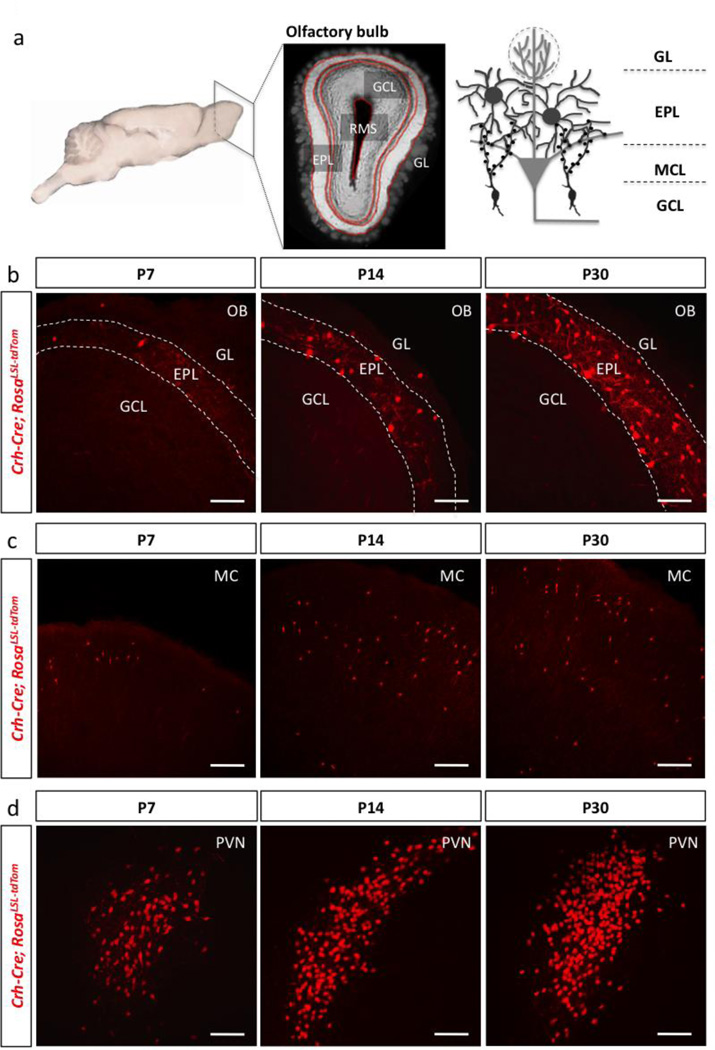
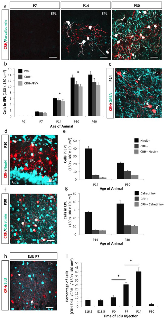
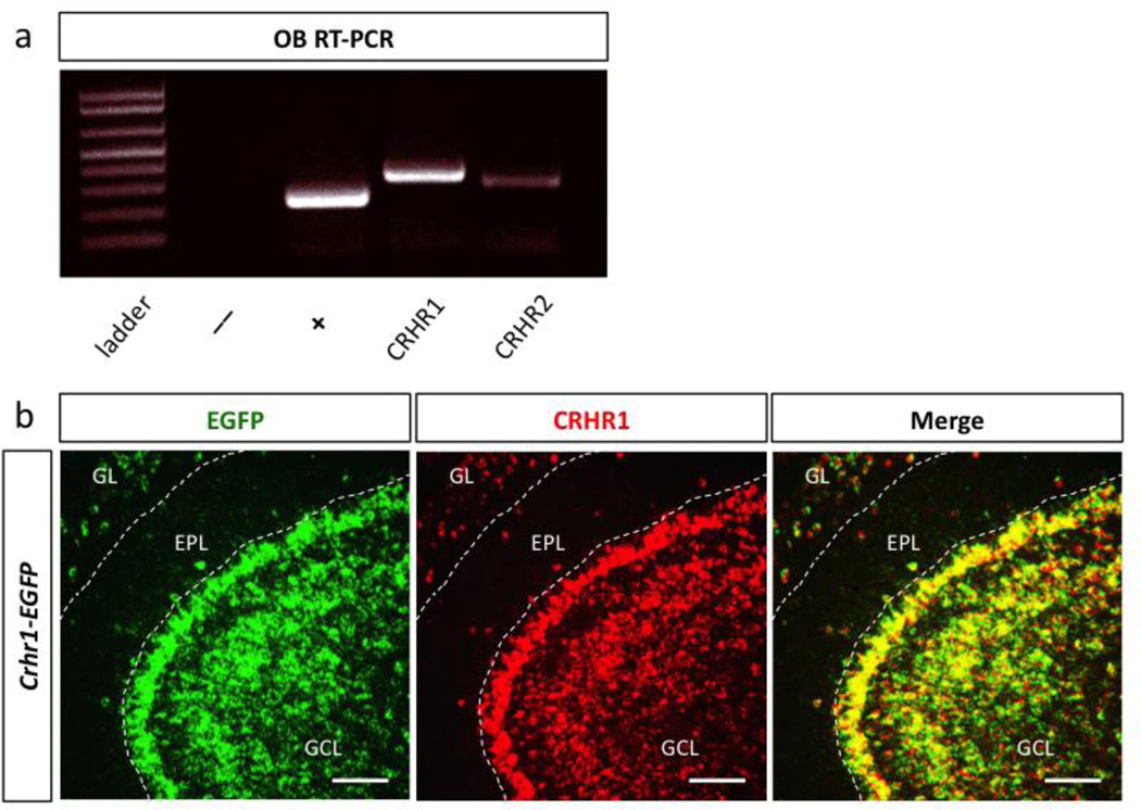
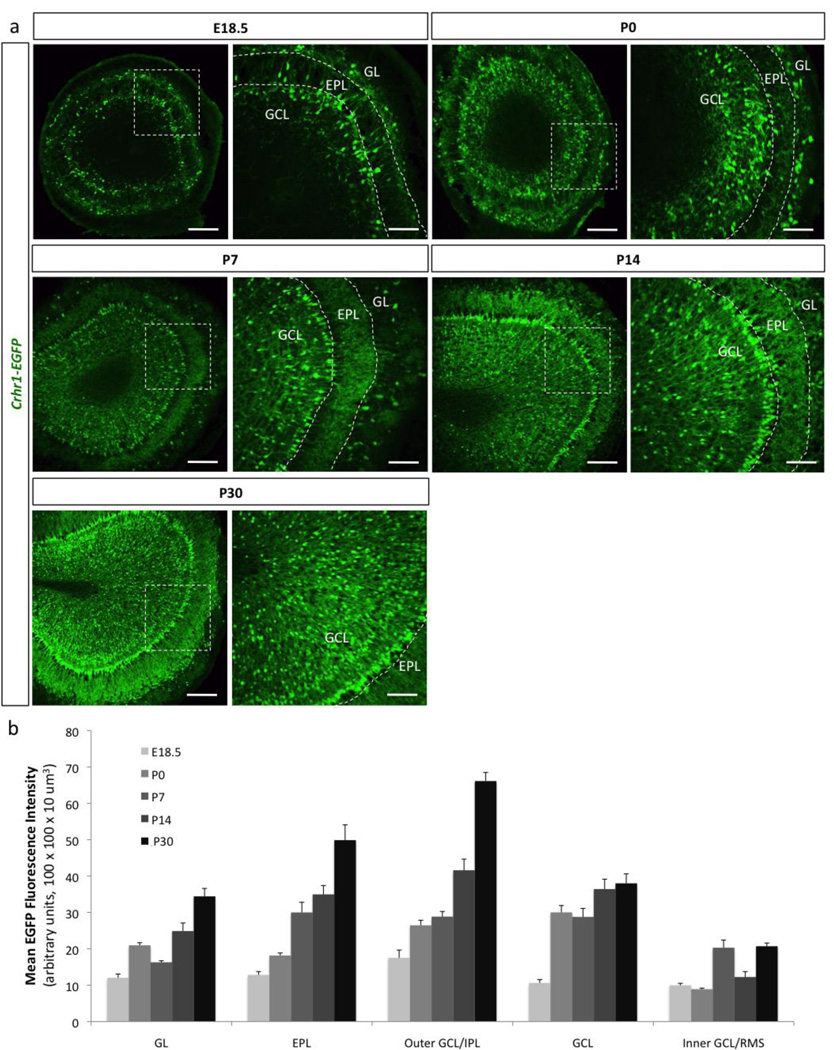
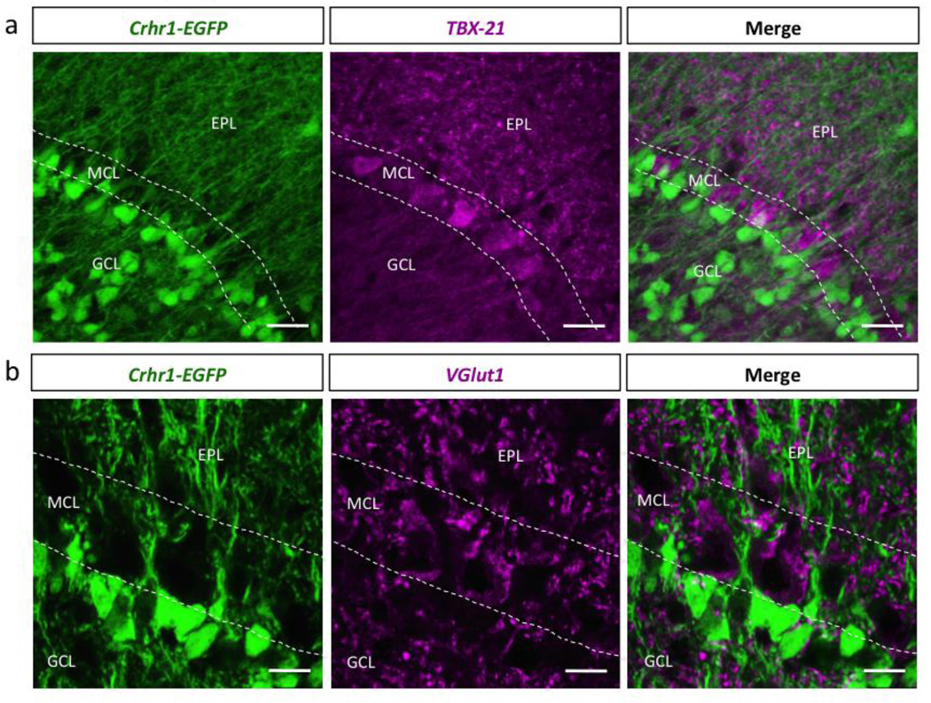
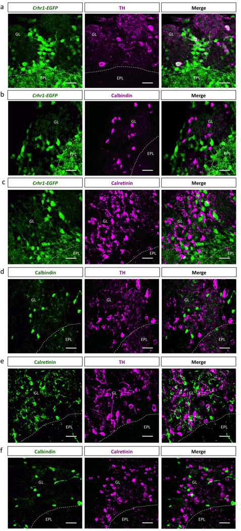
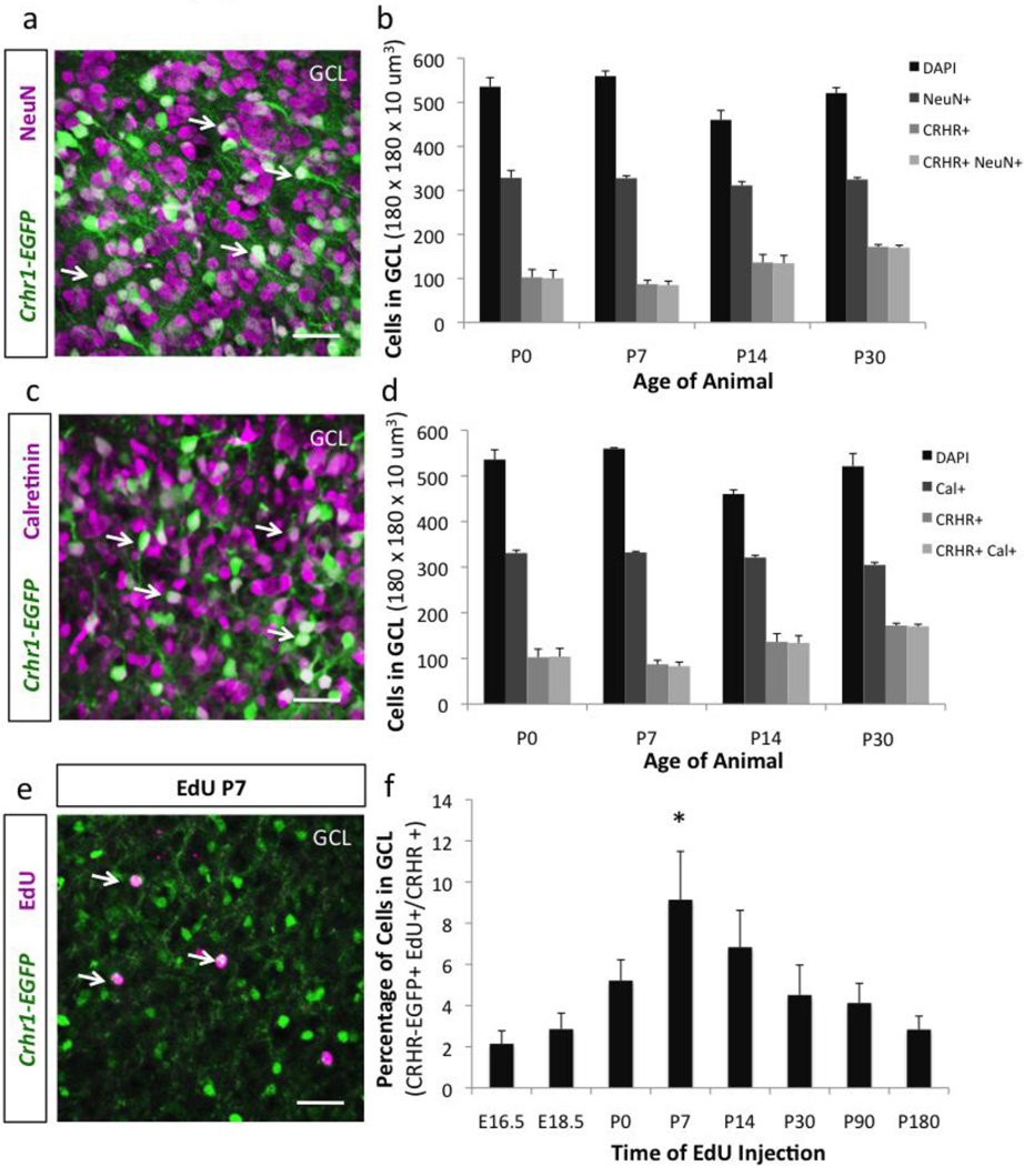
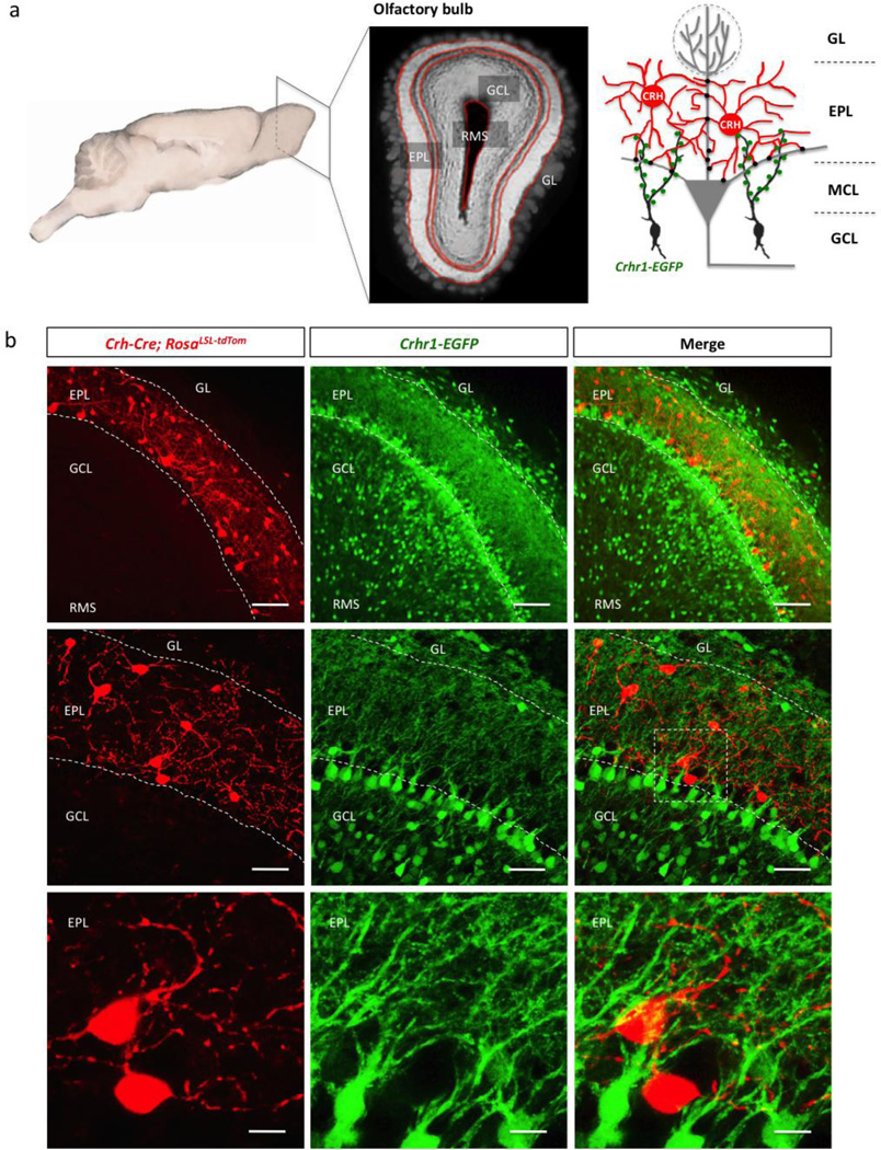
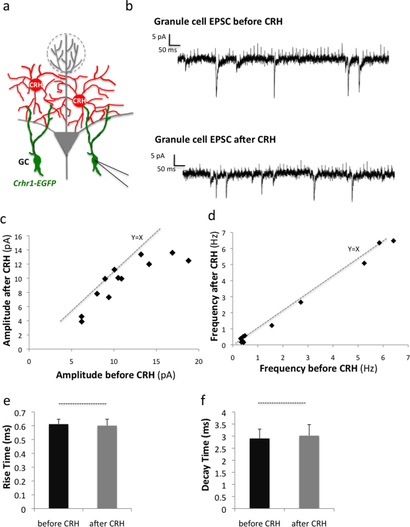
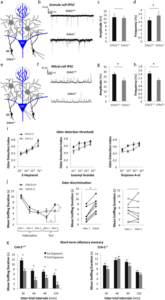
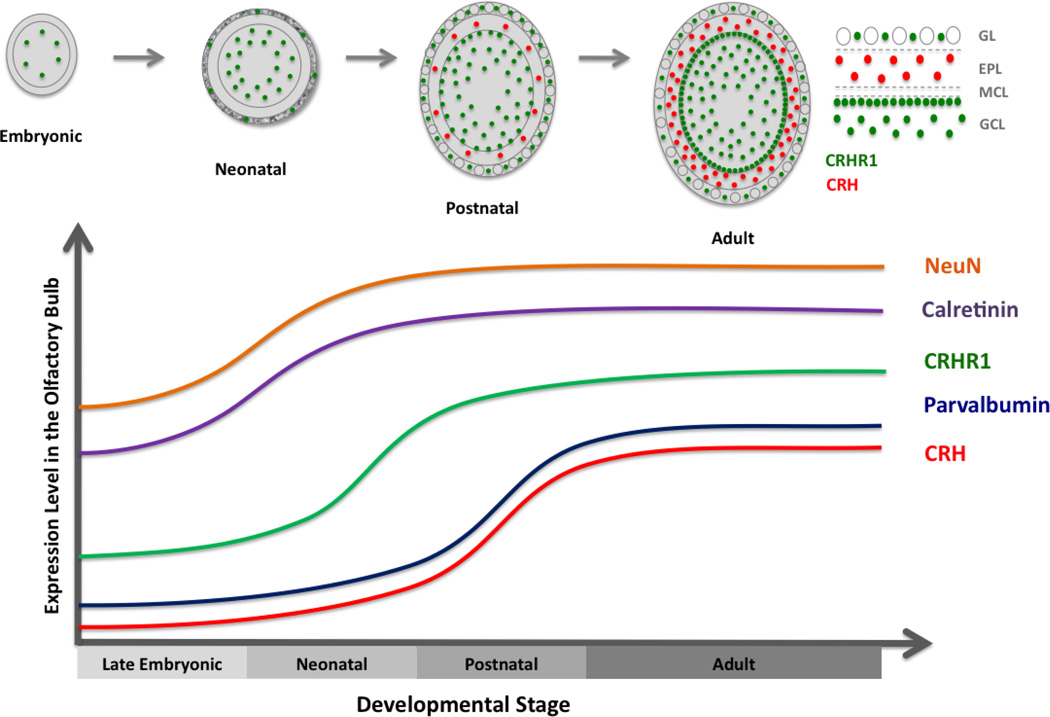
Neuropeptides play important physiological functions during distinct behaviors such as arousal, learning, memory, and reproduction. However, the role of local, extrahypothalamic neuropeptide signaling in shaping synapse formation and neuronal plasticity in the brain is not well understood. Here, we characterize the spatiotemporal expression profile of the neuropeptide corticotropin-releasing hormone (CRH) and its receptor CRHR1 in the mouse OB throughout development. We found that CRH-expressing interneurons are present in the external plexiform layer, that its cognate receptor is expressed by granule cells, and show that both CRH and CRHR1 expression enriches in the postnatal period when olfaction becomes important towards olfactory-related behaviors. Further, we provide electrophysiological evidence that CRHR1-expressing granule cells functionally respond to CRH ligand, and that the physiological circuitry of CRHR1 knockout mice is abnormal, leading to impaired olfactory behaviors. Together, these data suggest a physiologically relevant role for local CRH signaling towards shaping the neuronal circuitry within the mouse OB.

Keywords: CRF; CRFR1; CRH; CRHR1; EPL; GPCR; Granule cell; Neuropeptide; Olfactory.


Figures

Figure 1
Figure 1. CRH expression in olfactory bulb EPL interneurons is initiated during the postnatal period
(a) Simplified schematic of the olfactory bulb circuitry (RMS-rostral migratory stream, GCL-granule cell layer, EPL-external plexiform layer, MCL-mitral cell layer, GL-glomerular layer). Confocal image of the expression pattern of Crh-Cre; Rosalsl-tdTomato/+ mice at P7, P14, and P30 in the (b) olfactory bulb (scale bar 100 m), the (c) motor cortex (MC) (scale bar 150 m), and the (d) hypothalamic paraventricular nucleus (PVN) (scale bar 100 µm).
Figure 2
Figure 2. CRH+ EPL interneurons are predominantly generated postnatally
(a) Expression pattern of Crh-Cre; Rosalsl-tdTomato/+ mice at P7, P14, and P30 co-stained with the interneuron marker Parvalbumin (scale bar 30 µm). (b) Quantification of the expression of Parvalbumin and CRH+ interneurons in the EPL between P0 and P60. Confocal immunohistochemistry image of (c) GABA on Crh-Cre; Rosalsl-tdTomato/+ EPL tissue at P14 (scale bar 20 µm), and (d) NeuN at P30 (scale bar 30 µm). (e) Quantification of the expression of NeuN and CRH+ interneurons at P14 and P30. (f) Confocal image of Calretinin in CRH+ EPL interneurons at P30 (scale bar 40 µm). (g) Quantification of the expression of Calretinin and CRH+ interneurons at P14 and P30. (h) Example confocal image of EdU expression in CRH+ neurons pulsed at P7 (scale bar 40 m). (i) Quantification of the percentage of CRH+ EPL interneurons born between E16.5 and P30 (* p< 0.05). All data points represent averages +/− SEM.
Figure 3
Figure 3. The CRH receptor CRHR1 is enriched in the olfactory bulb
(a) Semi-quantitative RT-PCR of olfactory bulb cDNA amplifying CRHR1 and CRHR2 mRNA transcript (-ctrl RNA without reverse transcriptase, + ctrl GAPDH). (b) Double in situ hybridization image of EGFP and CRHR1 expression in Crhr1-EGFP BAC transgenic olfactory bulb slice (GCL-granule cell layer, EPL-external plexiform layer, GL-glomerular layer, scale bar 200 m).
Figure 4
Figure 4. Olfactory bulb CRHR1 expression enriches postnatally and into adulthood
(a) Olfactory bulb expression pattern of Crhr1-EGFP transgenic mice throughout development at E18.5, P0, P7, P14, and P30 (GL-glomerular layer, EPL-external plexiform layer, GCL-granule cell layer, scale bar 300 µm and 100 m for inserts, respectively). (b) Quantification of the layer-specific expression pattern of Crhr1-EGFP transgenic mice between E18.5 and P30. All data points represent averages +/− SEM.
Figure 5
Figure 5. CRHR1 is not expressed by olfactory bulb mitral cells
Immunohistochemistry confocal image of Crhr1-EGFP olfactory bulb tissue stained with (a) TBX-21 (GCL-granule cell layer, MCL-mitral cell layer, EPL-external plexiform layer, scale bar 50 m), and (b) VGlut1 (scale bar 20 m).
Figure 6
Figure 6. CRHR1 is expressed by a subset of dopaminergic periglomerular cells
Immunohistochemistry confocal images of Crhr1-EGFP olfactory bulb tissue stained with (a) Tyrosine Hydroxylase (TH) (EPL-external plexiform layer, GL-glomerular layer, scale bar 40 m), (b) Calbindin (scale bar 40 m), and (c) Calretinin (scale bar 40 m). Confocal images of wildtype olfactory bulb tissue stained with (d) Calbindin and TH (scale bar 40 m), (e) Calretinin and TH (scale bar 40 m), and (f) Calbindin and Calretinin (scale bar 40 m).
Figure 7
Figure 7. CRHR1+ granule cells are continuously generated throughout development with postnatal enrichment
(a) Confocal image showing immunodetection of NeuN in the granule cell layer (GCL) of Crhr1-EGFP olfactory bulb sections (scale bar 30 µm). (b) Quantification of the expression of NeuN and CRHR1+ interneurons in the GCL at P0, P7, P14, and P30. (c) Confocal image showing immunodetection of Calretinin in the GCL of Crhr1-EGFP olfactory bulb sections (scale bar 30 µm). (d) Quantification of the expression of Calretinin and CRHR1 interneurons in the GCL at P0, P7, P14, and P30. (e) Representative GCL confocal image of EdU-pulsed Crhr1-EGFP mice at P7 (scale bar 40 m). (f) Quantification of the percentage of CRHR1+ granule cells born between E16.5 and P180. All data points represent averages +/− SEM.
Figure 8
Figure 8. CRH and CRHR1 expressing interneurons are closely juxtaposed in the mouse olfactory bulb
(a) Simplified schematic of the mouse brain and olfactory bulb circuitry (GL-glomerular layer, EPL-external plexiform layer, MCL-mitral cell layer, GCL-granule cell layer, RMS-rostral migratory stream). (b) Confocal images of double reporter expression in the olfactory bulb of Crh-Cre; Rosalsl-tdTomato/+; Crhr1-EGFP mice. (scale bars 100, 50, and 10 m, respectively).
Figure 9
Figure 9. CRHR1-expressing granule cells show electrophysiological responses to CRH ligand
(a) Experimental scheme: Electrophysiological recordings are taken from CRHR1-expressing granule cells via patch-clamp before and after bath application of CRH ligand. (b) Representative traces showing EPSCs of recorded granule cells before and after CRH application. (c) Graph of EPSC amplitudes before and after CRH application. (d) Graph of EPSC frequency before and after CRH application. Quantification of the (e) rise time and (f) decay time of granule cell EPSCs. All data points represent mean +/− SEM (n= 12 cells per group from 3 mice).
Figure 10
Figure 10. Mice lacking CRHR1 show olfactory bulb circuit dysfunction and impaired olfactory behaviors
(a) Experimental scheme: Electrophysiological recordings are taken from granule cells in Crhr1−/− slices via patch-clamp. (b) Representative traces showing EPSCs from Crhr1+/+ and Crhr1−/− granule cells. Quantification of the (c) amplitudes and (d) frequencies of granule cell EPSCs (*p< 0.05, n=10 cells per group from 3 mice). (e) Experimental scheme: Electrophysiological recordings are taken from mitral cells in Crhr1−/− olfactory bulb slices. (f) Representative traces showing IPSCs from mitral cells of Crhr1+/+ and Crhr1−/− olfactory bulbs. Quantification of the (g) amplitudes and (h) frequencies of mitral cell IPSCs (*p< 0.05, n=10 cells per group from 3 mice). (i) Odor detection threshold of control and Crhr1−/− mice at concentrations from 10−7 to 10−3. (j) Odor discrimination and (k) short-term olfactory memory performance of Crhr1−/− mice compared to controls (* p<0.05). All behavior data points represent mean +/− SEM, n = 8 animals per group).
Figure 11
Figure 11. CRH and CRHR1 expression in the olfactory bulb become enriched in the postnatal period
During development, olfactory bulb CRHR1 expression appears first in the embryonic period and then gradually increases to reach a steady state in adulthood. CRH expression turns on postnatally and increases into early adulthood, remaining stable thereafter.

Similar articles

Cited by

References

    1. Abraham NM, Egger V, Shimshek DR, Renden R, Fukunaga I, Sprengel R, Seeburg PH, Klugmann M, Margrie TW, Schaefer AT, et al. Synaptic inhibition in the olfactory bulb accelerates odor discrimination in mice. Neuron. 2010;65:399–411. - PMC - PubMed
    1. Abrous DN, Koehl M, Le Moal M. Adult neurogenesis: from precursors to network and physiology. Physiol Rev. 2005;85:523–569. - PubMed
    1. Alvarez-Buylla A, Garcia-Verdugo JM. Neurogenesis in adult subventricular zone. J Neurosci. 2002;22:629–634. - PMC - PubMed
    1. Arenkiel BR, Hasegawa H, Yi JJ, Larsen RS, Wallace ML, Philpot BD, Wang F, Ehlers MD. Activity-induced remodeling of olfactory bulb microcircuits revealed by monosynaptic tracing. PLoS One. 2011;6:e29423. - PMC - PubMed
    1. Bale TL, Vale WW. CRF and CRF receptors: role in stress responsivity and other behaviors. Annu Rev Pharmacol Toxicol. 2004;44:525–557. - PubMed

Publication types

MeSH terms

Substances

LinkOut - more resources