Fluoroscopic views for safe insertion of lag screws into the posterior column of the acetabulum
- PMID: 25224878
- PMCID: PMC4169822
- DOI: 10.1186/1471-2474-15-303
Fluoroscopic views for safe insertion of lag screws into the posterior column of the acetabulum
Abstract
Background: Percutaneous lag screw fixation is an alternative treatment for non-displaced or minimally displaced posterior column fractures. This study aims to explore new fluoroscopic views of the acetabulum for safe percutaneous insertion of posterior column lag screws.
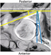
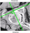
Methods: Axial computed tomography (CT) scans were taken of sixteen embalmed adult cadavers. The axial CT images at the level of the middle height of the acetabulum were selected. The angle (angle α) between the posterior cortex of the posterior column (PCPC) and the line intersecting the axial plane and the coronal plane, and the angle (angle β) between the medial wall and the line intersecting the axial plane and the sagittal plane were identified and measured. Tangential views of the PCPC and medial wall were obtained by referencing the measured angles. A lag screw was inserted into the posterior columns of the sixteen pelvic specimens under fluoroscopic guidance using an iliac oblique view and the two tangential views. CT scans were performed to evaluate the lag screw position. Axial CT images of 52 volunteers were obtained and the angles α and β were measured following the same methods used for the cadaveric specimens.
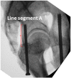
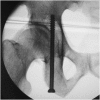
Results: The angles α and β for the specimens were 29.3 ± 2.8.1 and 8.1 ± 1.4 degrees, respectively. On the tangential view of the PCPC, the posterior cortex appears as a nearly straight line between the lesser and greater sciatic notches. On the tangential view of the medial wall, the medial wall appears as a distinct straight line. Using these radiographic images, the lag screws were inserted into the posterior columns of bony pelvic specimens. Screw placement was confirmed by CT, and found to be fully intraosseous in all cases without any cortical breaches. The angles α and β were 30.4 ± 4.1 and 9.2 ± 1.9 degrees for male volunteers and 28.5 ± 3.7 and 7.7 ± 1.8 degrees for female volunteers, significant difference in these angles between cadaveric specimens and human volunteers.
Conclusion: The tangential views of both the PCPC and medial wall can be obtained following the aforementioned methods The oblique iliac view and the two tangential views enable safe insertion of posterior column lag screws.
Figures


Similar articles
-
The study of plate-screw fixation in the posterior wall of acetabulum using computed tomography images.J Trauma. 2010 Aug;69(2):423-31. doi: 10.1097/TA.0b013e3181ca05f6. J Trauma. 2010. PMID: 20699753
-
Axial view of acetabular anterior column: a new X-ray projection of percutaneous screw placement.Arch Orthop Trauma Surg. 2015 Feb;135(2):187-192. doi: 10.1007/s00402-014-2127-0. Epub 2014 Dec 2. Arch Orthop Trauma Surg. 2015. PMID: 25450306
-
Study of anatomical parameters and intraoperative fluoroscopic techniques for transiliac crest anterograde lag screws fixation of the posterior column of the acetabulum.J Orthop Surg Res. 2023 Sep 18;18(1):697. doi: 10.1186/s13018-023-04208-3. J Orthop Surg Res. 2023. PMID: 37723587 Free PMC article.
-
[Percutaneous screw fixation in treatment of fractures of acetabular columns using computer-assisted imaging navigation system: experiment with cadaver model].Zhonghua Yi Xue Za Zhi. 2008 Jul 15;88(27):1900-4. Zhonghua Yi Xue Za Zhi. 2008. PMID: 19040003 Chinese.
-
[Digital study of the ideal position of lag screw internal fixation in the anterior column of the acetabulum].Zhongguo Xiu Fu Chong Jian Wai Ke Za Zhi. 2021 Jun 15;35(6):684-689. doi: 10.7507/1002-1892.202102002. Zhongguo Xiu Fu Chong Jian Wai Ke Za Zhi. 2021. PMID: 34142493 Free PMC article. Chinese.
Cited by
-
Virtual mapping of 260 three-dimensional hemipelvises to analyse gender-specific differences in minimally invasive retrograde lag screw placement in the posterior acetabular column using the anterior pelvic and midsagittal plane as reference.BMC Musculoskelet Disord. 2015 Sep 4;16:240. doi: 10.1186/s12891-015-0697-9. BMC Musculoskelet Disord. 2015. PMID: 26341003 Free PMC article.
-
Application of a three-dimensional virtual model to study the effect of fluoroscopic angle on infra-acetabular corridor parameters and screw insertion rates.J Orthop Surg Res. 2021 Sep 26;16(1):574. doi: 10.1186/s13018-021-02730-w. J Orthop Surg Res. 2021. PMID: 34565422 Free PMC article.
-
Antegrade Posterior Column Acetabulum Fracture Screw Fixation via Posterior Approach: A Biomechanical Comparative Study.Medicina (Kaunas). 2023 Jun 28;59(7):1214. doi: 10.3390/medicina59071214. Medicina (Kaunas). 2023. PMID: 37512026 Free PMC article.
-
The novel infra-pectineal buttress plates used for internal fixation of elderly quadrilateral surface involved acetabular fractures.Orthop Surg. 2022 Aug;14(8):1583-1592. doi: 10.1111/os.13327. Epub 2022 Jun 15. Orthop Surg. 2022. PMID: 35706090 Free PMC article.
-
Infra-acetabular screw exited between ischial tuberosity and ischial spine is more suitable for Asian population: a 3D morphometric study.BMC Musculoskelet Disord. 2020 Nov 28;21(1):787. doi: 10.1186/s12891-020-03802-4. BMC Musculoskelet Disord. 2020. PMID: 33248460 Free PMC article.
References
-
- Sanders MB, Starr AJ, Reinert C. Percutaneous screw fixation of acetabular fractures in elderly patients. Curr Opin Orthop. 2006;17(1):17–24.
Pre-publication history
-
- The pre-publication history for this paper can be accessed here:http://www.biomedcentral.com/1471-2474/15/303/prepub
Publication types
MeSH terms
LinkOut - more resources
Full Text Sources
Other Literature Sources

