. 2014 Dec;18(12):2488-98.
doi: 10.1111/jcmm.12389. Epub 2014 Sep 16.

Delayed cutaneous wound closure in HO-2 deficient mice despite normal HO-1 expression

Affiliations

Delayed cutaneous wound closure in HO-2 deficient mice despite normal HO-1 expression

Ditte M S Lundvig et al. J Cell Mol Med. 2014 Dec.

Abstract

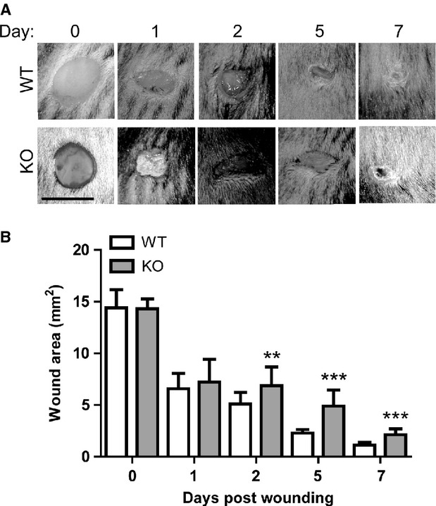
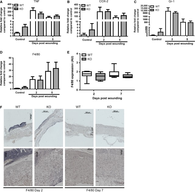
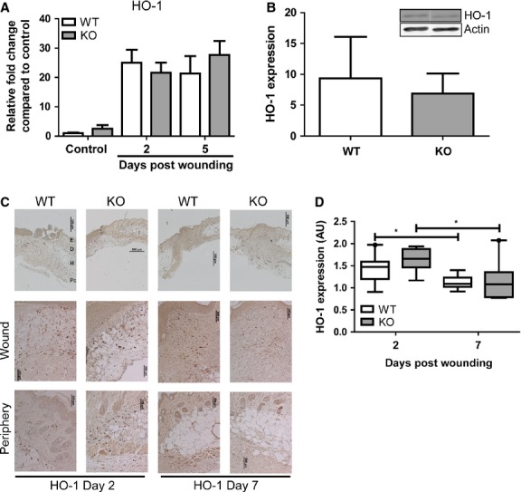
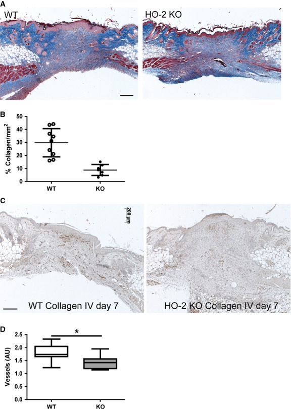
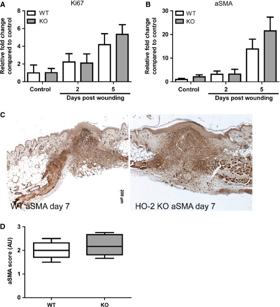
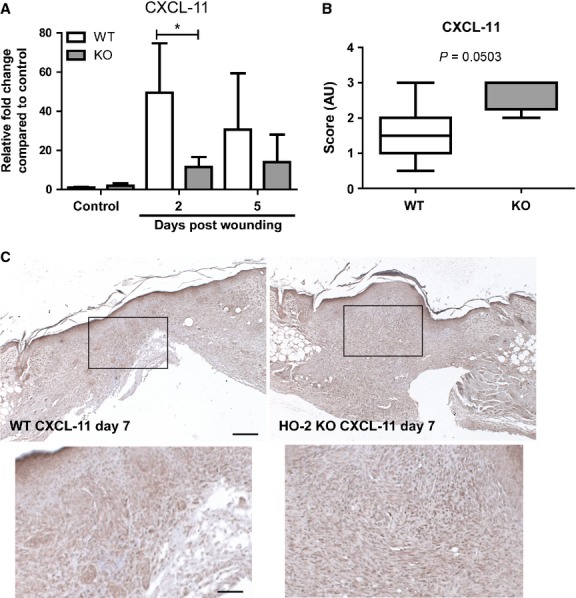
Impaired wound healing can lead to scarring, and aesthetical and functional problems. The cytoprotective haem oxygenase (HO) enzymes degrade haem into iron, biliverdin and carbon monoxide. HO-1 deficient mice suffer from chronic inflammatory stress and delayed cutaneous wound healing, while corneal wound healing in HO-2 deficient mice is impaired with exorbitant inflammation and absence of HO-1 expression. This study addresses the role of HO-2 in cutaneous excisional wound healing using HO-2 knockout (KO) mice. Here, we show that HO-2 deficiency also delays cutaneous wound closure compared to WT controls. In addition, we detected reduced collagen deposition and vessel density in the wounds of HO-2 KO mice compared to WT controls. Surprisingly, wound closure in HO-2 KO mice was accompanied by an inflammatory response comparable to WT mice. HO-1 induction in HO-2 deficient skin was also similar to WT controls and may explain this protection against exaggerated cutaneous inflammation but not the delayed wound closure. Proliferation and myofibroblast differentiation were similar in both two genotypes. Next, we screened for candidate genes to explain the observed delayed wound closure, and detected delayed gene and protein expression profiles of the chemokine (C-X-C) ligand-11 (CXCL-11) in wounds of HO-2 KO mice. Abnormal regulation of CXCL-11 has been linked to delayed wound healing and disturbed angiogenesis. However, whether aberrant CXCL-11 expression in HO-2 KO mice is caused by or is causing delayed wound healing needs to be further investigated.

Keywords: haem oxygenase; skin; wound healing.


Figures

Fig. 1
Fig. 1
Slower cutaneous wound closure in HO-2 KO after excisional wounding. (A) Wound closure in WT (upper panel) and HO-2 KO (lower panel) mice in time; bar, 5 mm. (B) Wound area (mm2) reduction in WT (white bars) and HO-2 KO (grey bars) mice in time presented as mean ± SD. **P < 0.01, ***P < 0.001.
Fig. 2
Fig. 2
HO-2 KO mice demonstrate a normal inflammatory response after wounding. Gene transcript levels of pro-inflammatory proteins (A) TNF and (B) COX-2 and inflammatory cell markers (C) Gr-1, and (D) F4/80 in WT (white bars) and HO-2 KO (grey bars) mice in time presented as mean ± SD. Controls are tissue biopsies collected at day 0, and data were normalized to WT mean day 0. (E) Box-and-whisker plot with 10–90 percentiles of semi-quantitative assessment of F4/80 immunoreactivity in (F). (F) F4/80 immunoreactivity in representative wound sections of WT and HO-2 KO mice at day 2 and 7 after wounding. Anatomical indications by E, epidermis; D, dermis; H, hypodermis; Pc, panniculus carnosus; bars, 500 μm (upper panel), 100 μm (lower panel).
Fig. 3
Fig. 3
HO-2 KO mice induce cutaneous HO-1 expression after wounding. (A) HO-1 gene transcript levels in WT (white bars) and HO-2 KO (grey bars) mice in time as represented as mean ± SD. Controls represent biopsies collected at day 0, and data were normalized to WT mean day 0. (B) Western blot (insert) of cutaneous HO-1 expression in unwounded skin in WT (white bar) and HO-2 KO (grey bar) mice. Band intensity was normalized to housekeeping protein β-actin. Data are presented as mean ± SD. (C) HO-1 immunoreactivity in representative wound sections of WT and HO-2 KO mice at day 2 and day 7 after wounding. Anatomical indications by E, epidermis; D, dermis; H, hypodermis; Pc, panniculus carnosus; bars, 500 μm (upper panel), 100 μm (wound, periphery). (D) Semi-quantitative scores of HO-1 immunoreactivity in (C) presented as box-and-whisker plot with 10–90 percentiles. *P < 0.05.
Fig. 4
Fig. 4
Reduced collagen deposition and vessel density in HO-2 KO mice. (A) Representative images of AZAN stained wound sections of WT and HO-2 KO mice at day 7 after wounding; bar, 200 μm. (B) Collagen deposition in WT (open circles) and HO-2 KO (closed circles) mice at day 7 after wounding. (C) Representative images of collagen IV immunoreactivity, a common blood vessel marker, in WT and HO-2 KO mice at day 7 after wounding; bar, 200 μm. (D) Semi-quantitative scoring of high-power fields of (C). Data are represented as box-and-whisker plot with 10–90 percentiles. *P < 0.05, ***P < 0.001.
Fig. 5
Fig. 5
Myofibroblast differentiation occurs in HO-2 KO mice. Ki67 (A) and ACTA2 (B) gene transcript levels in WT (white bars) and HO-2 KO (grey bars) mice in time as represented as mean ± SD. Controls represent biopsies collected at day 0, and data were normalized to WT mean day 0. (C) Representative images of αSMA immunoreactivity in wounds of WT and HO-2 KO mice at day 7 after wounding; bar, 200 μm. (D) Semi-quantitative scoring of αSMA in (C) presented as box-and-whisker plot with 10–90 percentiles.
Fig. 6
Fig. 6
Different CXCL-11 expression in HO-2 KO and WT mice after injury. (A) CXCL-11 gene transcript levels in WT (white bars) and HO-2 KO (grey bars) mice in time presented as mean ± SD. Controls represent biopsies collected at day 0, and data were normalized to WT mean day 0. (B) Semi-quantitative scoring of CXCL-11 immunoreactivity in (C) of WT (white) and HO-2 KO (grey) wounds presented as box-and-whisker plot with 10–90 percentiles. *P < 0.05. (C) Representative images of CXCL-11 immunoreactivity in wound tissue of WT and HO-2 KO mice at day 7 after wounding; bar, 200 μm. Insert represent magnified boxed area; bar, 70 μm.

Similar articles

Cited by

References

    1. Martin P. Wound healing–aiming for perfect skin regeneration. Science. 1997;276:75–81. - PubMed
    1. Buganza Tepole A, Kuhl E. Systems-based approaches toward wound healing. Pediatr Res. 2013;73:553–63. - PMC - PubMed
    1. Sidgwick GP, Bayat A. Extracellular matrix molecules implicated in hypertrophic and keloid scarring. J Eur Acad Dermatol Venereol. 2012;26:141–52. - PubMed
    1. Grice EA, Segre JA. Interaction of the microbiome with the innate immune response in chronic wounds. Adv Exp Med Biol. 2012;946:55–68. - PMC - PubMed
    1. Otterbein LE, Choi AM. Heme oxygenase: colors of defense against cellular stress. Am J Physiol Lung Cell Mol Physiol. 2000;279:L1029–37. - PubMed

Publication types

MeSH terms