. 2014 Dec;64(6):1368-1375.
doi: 10.1161/HYPERTENSIONAHA.114.03743. Epub 2014 Sep 15.

Angiotensin II mediates angiotensin converting enzyme type 2 internalization and degradation through an angiotensin II type I receptor-dependent mechanism

Affiliations

Angiotensin II mediates angiotensin converting enzyme type 2 internalization and degradation through an angiotensin II type I receptor-dependent mechanism

Matthew R Deshotels et al. Hypertension. 2014 Dec.

Erratum in

  • Hypertension. 2014 Dec;64(6):e8. Sriramula, Srinivas [added]

Abstract

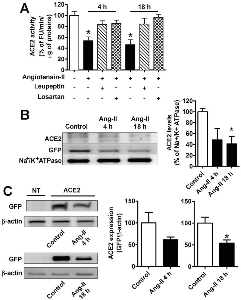
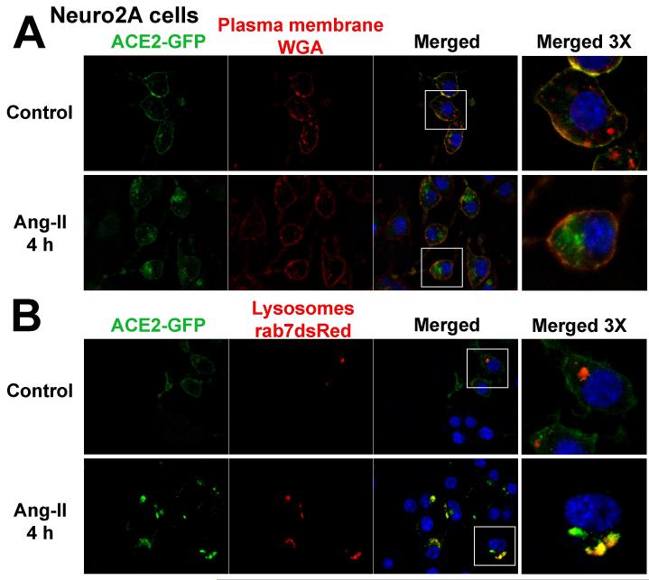
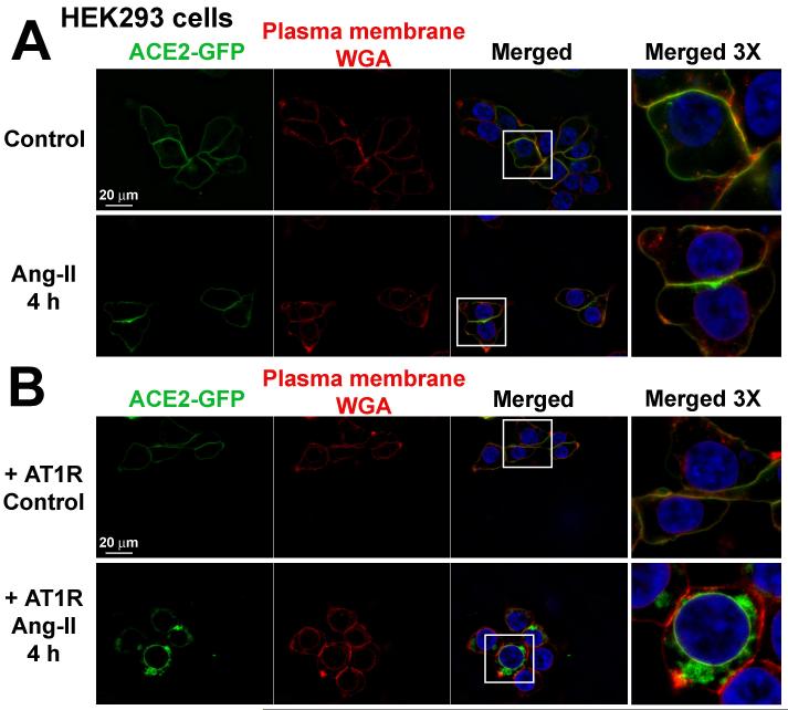
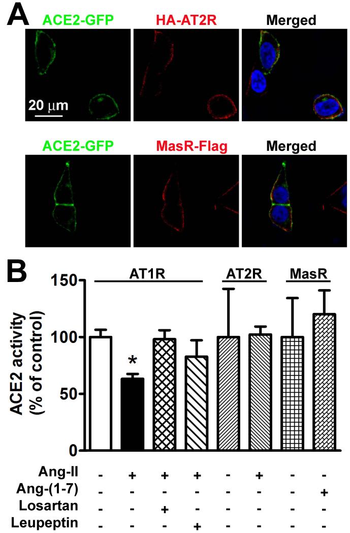
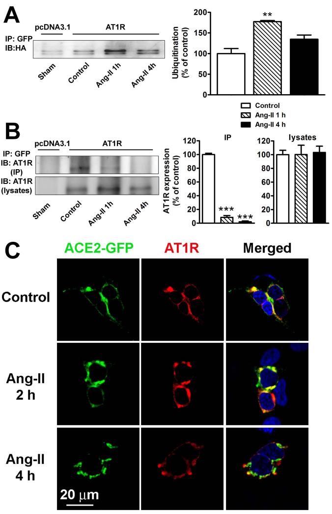
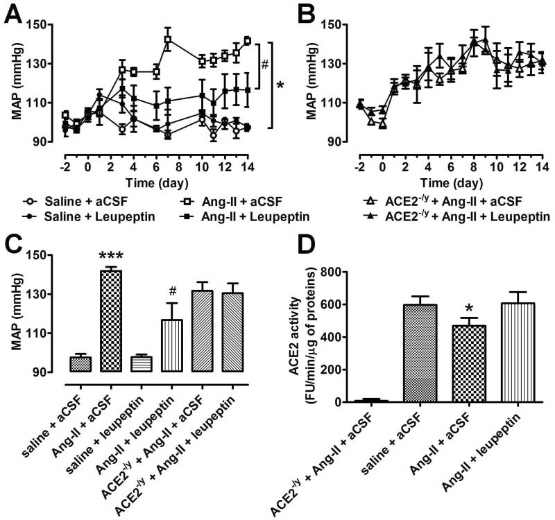
Angiotensin-converting enzyme type 2 (ACE2) is a pivotal component of the renin-angiotensin system, promoting the conversion of angiotensin II (Ang-II) to Ang-(1-7). We previously reported that decreased ACE2 expression and activity contributes to the development of Ang-II-mediated hypertension in mice. The present study aimed to investigate the mechanisms involved in ACE2 downregulation during neurogenic hypertension. In ACE2-transfected Neuro-2A cells, Ang-II treatment resulted in a significant attenuation of ACE2 enzymatic activity. Examination of the subcellular localization of ACE2 revealed that Ang-II treatment leads to ACE2 internalization and degradation into lysosomes. These effects were prevented by both the Ang-II type 1 receptor (AT1R) blocker losartan and the lysosomal inhibitor leupeptin. In contrast, in HEK293T cells, which lack endogenous AT1R, Ang-II failed to promote ACE2 internalization. Moreover, this effect could be induced after AT1R transfection. Furthermore, coimmunoprecipitation experiments demonstrated that AT1R and ACE2 form complexes, and these interactions were decreased by Ang-II treatment, which also enhanced ACE2 ubiquitination. In contrast, ACE2 activity was not changed by transfection of AT2 or Mas receptors. In vivo, Ang-II-mediated hypertension was blunted by chronic infusion of leupeptin in wildtype C57Bl/6, but not in ACE2 knockout mice. Overall, this is the first demonstration that elevated Ang-II levels reduce ACE2 expression and activity by stimulation of lysosomal degradation through an AT1R-dependent mechanism.

Keywords: autonomic nervous system; hypertension; proteasome endopeptidase complex; renin-angiotensin system.


Figures

Figure 1
Figure 1
A. ACE2 activity in transfected Neuro-2A cells in control (white bar), or after treatment with Ang-II alone (100 nM, black bars), or Ang-II and losartan (1 μM, horizontal hatched bars), or Ang-II and leupeptin (100 μM, vertical hatched bars) after 4 h (middle columns) or 18 h (right columns). ACE2 activity was determined as described in Experimental Procedures and the results were expressed as % from the activity measured in control cells (100% corresponds to 449 ±51 FU/min/μg of proteins). n=12 from 5 independent transfections, *−indicates P<0.05. B. Plasma membrane levels of ACE2 in control conditions and after treatment with 100 nM Ang-II for 4 and 18 h. Neuro-2A cells were transfected with 2 μg/well ACE2-GFP in 6-well plates and 48 h later the plasma membrane proteins were isolated by biotinylation, as described in Methods section. Subsequently, ACE2 levels were determined by western blot using ACE2 (top) or GFP (middle) antibodies. Na+/K+ ATP-ase (bottom) was used as a loading control. The summary data of three independent experiments are shown in the bottom panel. *−indicates P<0.05. C. Total cellular levels of ACE2-GFP in Neuro-2A cells in non-treated controls and after Ang-II (100 nM) treatment for 18 h. Similar results were obtained in two other independent experiments. FU: Fluorescence units.
Figure 2
Figure 2
Subcellular localization of ACE2-GFP in Neuro-2A cells treated with WGA (wheat germ agglutinin) for plasma membrane localization (A) and co-transfected with Rab7-dsRed for labeling of lysosomal compartments (B). Cells were processed as described in the Methods section. These images are representative of 12 different coverslips from 5 independent transfections. The far right panels represent 3 times magnification of the white boxes in the merged column.
Figure 3
Figure 3
Subcellular localization of ACE2-GFP in HEK293T cells co-transfected with pcDNA3.1 (A) or AT1R (B). The cells were treated with wheat germ agglutinin (WGA) for plasma membrane staining as described in the Methods section. These images are representative of 12 different coverslips from 4 independent transfections. The far right panels represent 3 times magnification of the white boxes in the merged column.
Figure 4
Figure 4
A. AT2R (top panel) and MasR (bottom panel) are localized at the plasma membrane in transfected HEK293T cells, but are not co-localized with ACE2. These images are representative of 6 different coverslips from 3 independent transfections. B. ACE2 activity in HEK293T cells co-transfected with AT1R (left columns), AT2R (middle columns) and MasR (right columns) in presence of different treatments indicated under each bar. n=6 from 2 independent transfections. *−indicates P<0.05.
Figure 5
Figure 5
A. Ang-II stimulates ACE2 ubiquitination. HEK293T cells were transfected in 10 cm2 plates with 10 μg ACE2-GFP, 10 μg HA-ubiquitin, and either 10 μg pcDNA3.1 (1st lane) or 10 μg AT1R (lanes 2-4). Cells were serum starved for 24 h and treated with Ang-II (100 nM) for the indicated time periods. Cells were then lysed and ACE2 interacting proteins were pulled down by treatment with a GFP antibody (2 μg/mg). Cell lysates were separated by 10% SDS-PAGE and ubiquitin levels were revealed by western blotting using anti-HA antibody. The experiment shown is representative from 3 independent transfections. **−indicates P<0.01 B. ACE2 interaction with AT1R in HEK293T cells determined by co-immunoprecipitation. Cells were transfected in 10 cm2 plates with 10 μg ACE2-GFP and 10 μg pcDNA3.1 (lane 1) or 10 μg AT1R (lanes 2-4). Cells were serum starved for 24 hours and treated with Ang-II (100 nM) for the indicated time periods. Following lysis, ACE2 interacting proteins were pulled down by treatment with a GFP antibody (2 μg/mg). AT1R was detected in immunoprecipitates (top panel) and lysates (bottom panel) using an anti-AT1R antibody (SC-579). Similar results were obtained in 2 other experiments. ***−indicates P<0.001 C. ACE2 and AT1R localization in HEK293T cells in control conditions (top panel), or after 2 h (middle panel) and 4 h (bottom panel) treatment with Ang-II (100 nM).
Figure 6
Figure 6
A. The effects of Ang-II and leupeptin on blood pressure in wild type mice (n=6-8 animals per group). B. The effects of Ang-II and leupeptin on blood pressure in ACE2 KO mice (n=6-8 animals per group). C. Average MAP following 2 weeks of treatment in all 6 groups. ***−indicates P<0.001 between saline + aCSF vs. Ang-II + aCSF; #-indicates P<0.05 between Ang-II + leupeptin vs. Ang-II + aCSF. D. ACE2 activity in the hypothalamus of animals used in A and B. n=4-5 per group, *−indicates P<0.05 between Ang-II + aCSF vs. saline + aCSF.

Comment in

References

    1. Lazartigues E, Feng Y, Lavoie JL. The two fACEs of the tissue renin-angiotensin systems: implication in cardiovascular diseases. Curr Pharm Des. 2007;13:1231–1245. - PubMed
    1. Xu P, Sriramula S, Lazartigues E. ACE2/Ang-(1-7)/Mas pathway in the brain: The Axis of Good. Am J Physiology - Regul Integr Comp Physiol. 2011;300:R804–817. - PMC - PubMed
    1. Xia H, Feng Y, Obr TD, Hickman PJ, Lazartigues E. Angiotensin II Type 1 Receptor-Mediated Reduction of Angiotensin-Converting Enzyme 2 Activity in the Brain Impairs Baroreflex Function in Hypertensive Mice. Hypertension. 2009;53:210–216. - PMC - PubMed
    1. Feng Y, Hans C, McIlwain E, Varner KJ, Lazartigues E. Angiotensin-Converting Enzyme 2 Over-Expression in the Central Nervous System Reduces Angiotensin-II-Mediated Cardiac Hypertrophy. PLos ONE. 2012;7:e48910. - PMC - PubMed
    1. Xia H, Sriramula S, Chhabra K, Lazartigues E. Brain ACE2 Shedding Contributes to the Development of Neurogenic Hypertension. Circ Res. 2013;113:1087–1096. - PMC - PubMed

Publication types

Substances