Review
. 2014 Sep 15;21(10):527-33.
doi: 10.1101/lm.034355.114. Print 2014 Oct.

Higher brain functions served by the lowly rodent primary visual cortex

Affiliations
Review

Higher brain functions served by the lowly rodent primary visual cortex

Jeffrey P Gavornik et al. Learn Mem. .

Abstract

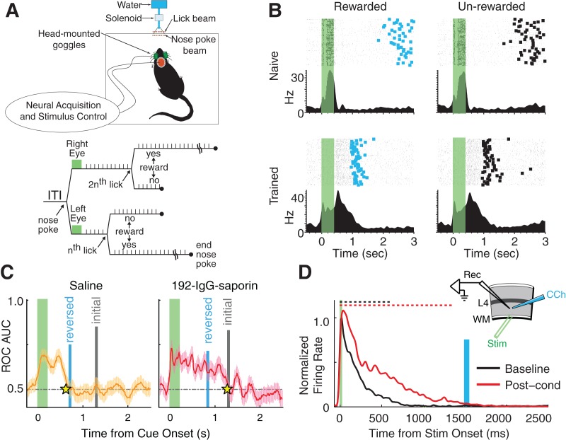
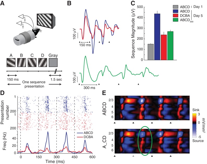
It has been more than 50 years since the first description of ocular dominance plasticity--the profound modification of primary visual cortex (V1) following temporary monocular deprivation. This discovery immediately attracted the intense interest of neurobiologists focused on the general question of how experience and deprivation modify the brain as a potential substrate for learning and memory. The pace of discovery has quickened considerably in recent years as mice have become the preferred species to study visual cortical plasticity, and new studies have overturned the dogma that primary sensory cortex is immutable after a developmental critical period. Recent work has shown that, in addition to ocular dominance plasticity, adult visual cortex exhibits several forms of response modification previously considered the exclusive province of higher cortical areas. These "higher brain functions" include neural reports of stimulus familiarity, reward-timing prediction, and spatiotemporal sequence learning. Primary visual cortex can no longer be viewed as a simple visual feature detector with static properties determined during early development. Rodent V1 is a rich and dynamic cortical area in which functions normally associated only with "higher" brain regions can be studied at the mechanistic level.


Figures

Figure 1.
Figure 1.
Stimulus-specific response potentiation (SRP). (A) Visual-evoked potentials (VEPs) are recorded from both hemispheres of head-fixed, awake, male C57BL/6 mice and driven by full-field, high-contrast, phase-reversing sinusoidal grating stimuli. Electrodes are positioned in Layer 4 of the binocular region of V1. (B) The trough-to-peak magnitude of average VEPs evoked by a stimulus oriented at X° (blue) potentiates over multiple viewings (Days 1–5). This potentiation is selective for the specific stimulus, demonstrated by the fact that VEPs evoked by novel orientations (on Days 5 and 9, offset in 45° increments from X°, red and green) have magnitudes equivalent to the Day 1 magnitude for X° and also potentiate across repeated presentations. (Insets) Representative VEP waveforms at indicated time points (scale bars, 100 μV × 50 msec). (All data adapted from Frenkel et al. 2006).
Figure 2.
Figure 2.
Reward-modulated interval timing learned in V1. (A) Schematic of the in vivo experimental design and trial flow from Shuler and Bear (2006). Light flashes were delivered to either the left or right eye following nose pokes and the number of licks (i.e., time) required before water-reward delivery depended on which eye was stimulated. (B) In naïve animals (top row), the duration of evoked responses (raster plots over PSTH) matched the stimulus duration (400 msec, indicated by green rectangle). After training, evoked activity persisted until the time of reward delivery (blue rectangles, left column), even on unrewarded trials (right column, time when reward would have been given indicated by thick black rectangles). (C) In Chubykin et al. (2013), rats were trained with a long interval between stimulus and reward and then treated with local injections of either IgG-saporin (to lesion cholinergic projections) or saline (control) in V1. The rats were then retrained with a short interval. Whereas the control animals were able to learn the new shortened interval, those with lesions continue to report the original timing. In both panels, the gray and blue vertical lines show the average reward-delivery times before and after lesioning, the star indicates the average reported time post-lesion, and neural interval reports are determined by a receiver operating characteristic (ROC) threshold crossing (see Chubykin et al. 2013 for details). (D) Repeated delivery of carbachol (a cholinergic agonist) after electric white matter stimulation produced a period of evoked spiking in mouse V1 slices that matched the interval between the two stimuli (electrical stimulation at time 0, CCh delivery indicated by blue rectangle, dashed lines show average scored response durations). (Data adapted from Shuler and Bear 2006 [A,B], and Chubykin et al. 2013 [C,D].)
Figure 3.
Figure 3.
Learned spatiotemporal sequence representations in V1. (A) Four-element sequences with a fixed timing (150 msec per element followed by 1.5 sec of gray screen) were shown to head-fixed male C57BL/6 mice repeatedly over 5 d. (B,C) The average sequence-evoked response (recorded in Layer 4) potentiated over days. On the fifth day, evoked field potentials were larger when the sequence elements were presented with the trained order and timing (ABCD, blue, triangles indicate the onset of each sequence element) than when they were reversed (DCBA, red) or slowed down (ABCD300, green, subscript indicates that each element is on screen for 300 msec). (D) Extracellular recordings show that the trained sequence ABCD elicits more spiking than the reversed sequence DCBA (color-coded spike rasters over PSTHs, dashed black lines show element onset times). (E) Current source density analysis of laminar field recordings (16 electrodes spaced every 50 μm from surface to white matter) show that the characteristic source–sink activation pattern evoked across the cortical layers by the sequence ABCD persists even when element B is not shown (A_CD, second element replaced with a gray screen, predictive response at the time that a stimulus was expected to be seen is highlighted with a green oval). (All data adapted from Gavornik and Bear 2014.)

References

    1. Alink A, Schwiedrzik CM, Kohler A, Singer W, Muckli L 2010. Stimulus predictability reduces responses in primary visual cortex. J Neurosci 30: 2960–2966 - PMC - PubMed
    1. Ayaz A, Saleem AB, Scholvinck ML, Carandini M 2013. Locomotion controls spatial integration in mouse visual cortex. Curr Biol 23: 890–894 - PMC - PubMed
    1. Bach-y-Rita P, Kercel SW 2003. Sensory substitution and the human-machine interface. Trends Cogn Sci 7: 541–546 - PubMed
    1. Balas B, Sinha P 2008. Observing object motion induces increased generalization and sensitivity. Perception 37: 1160–1174 - PubMed
    1. Balas BJ, Sinha P 2009. The role of sequence order in determining view canonicality for novel wire-frame objects. Atten Percept Psychophys 71: 712–723 - PubMed

Publication types

LinkOut - more resources