. 2014 Sep 22;211(10):1993-2011.
doi: 10.1084/jem.20132496. Epub 2014 Sep 15.

The Src family kinases Hck, Fgr, and Lyn are critical for the generation of the in vivo inflammatory environment without a direct role in leukocyte recruitment

Affiliations

The Src family kinases Hck, Fgr, and Lyn are critical for the generation of the in vivo inflammatory environment without a direct role in leukocyte recruitment

Miklós Kovács et al. J Exp Med. .

Abstract

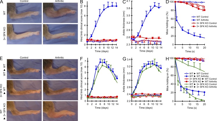
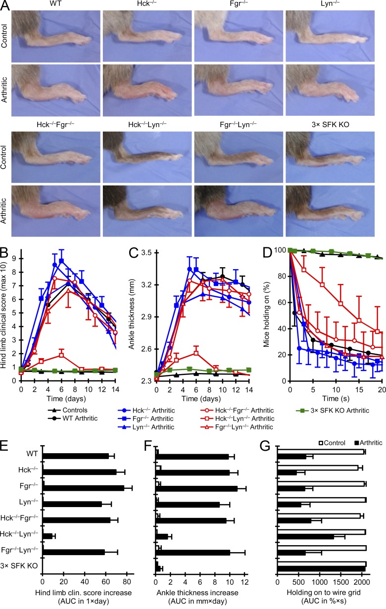
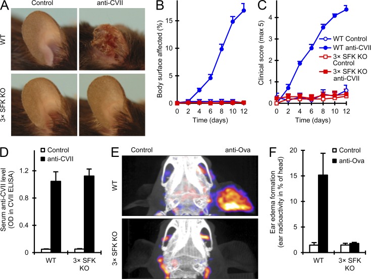
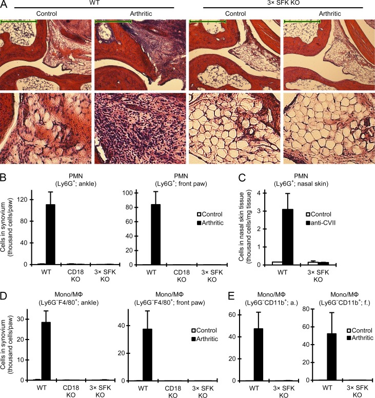
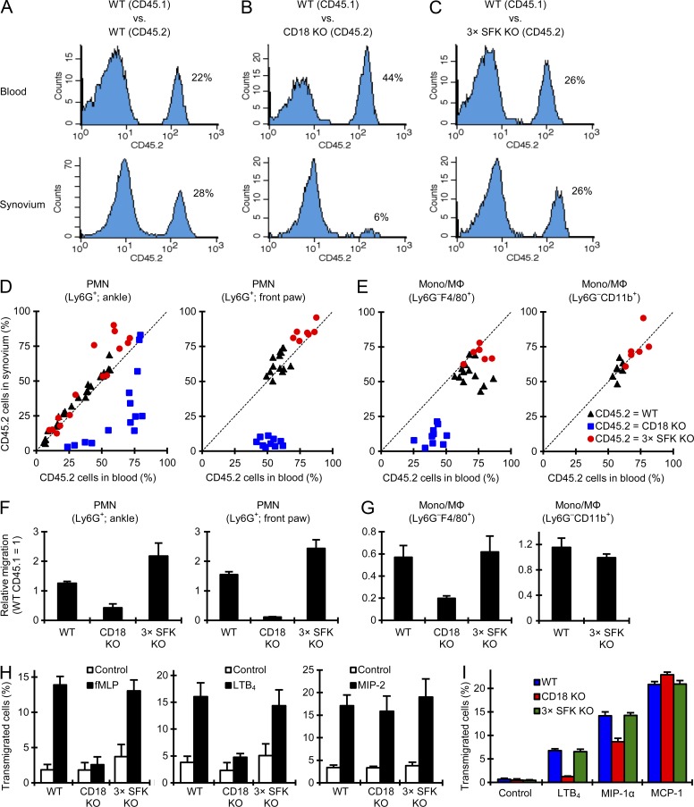
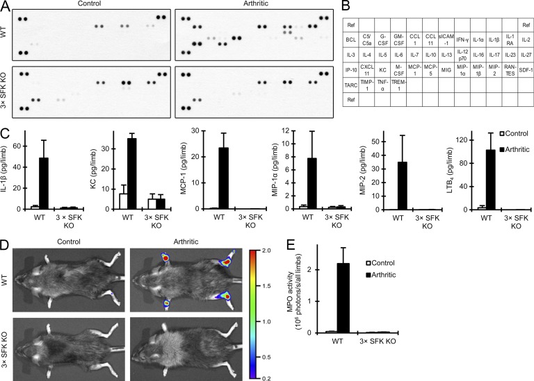
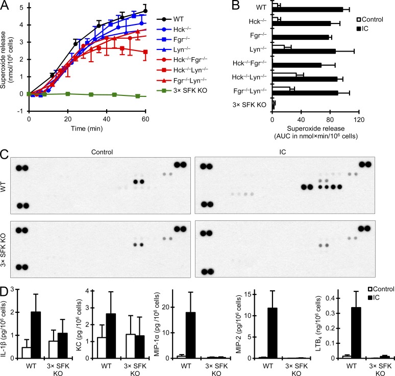
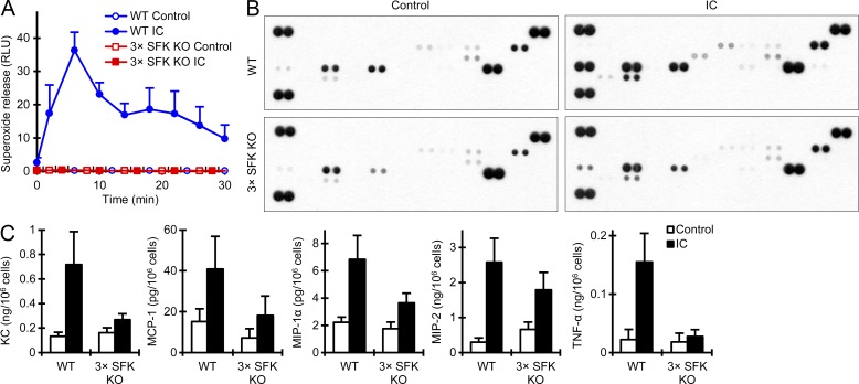
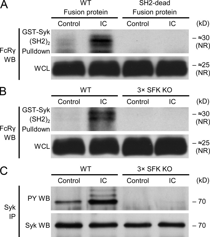
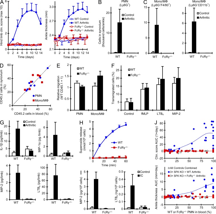
Although Src family kinases participate in leukocyte function in vitro, such as integrin signal transduction, their role in inflammation in vivo is poorly understood. We show that Src family kinases play a critical role in myeloid cell-mediated in vivo inflammatory reactions. Mice lacking the Src family kinases Hck, Fgr, and Lyn in the hematopoietic compartment were completely protected from autoantibody-induced arthritis and skin blistering disease, as well as from the reverse passive Arthus reaction, with functional overlap between the three kinases. Though the overall phenotype resembled the leukocyte recruitment defect observed in β2 integrin-deficient (CD18(-/-)) mice, Hck(-/-)Fgr(-/-)Lyn(-/-) neutrophils and monocytes/macrophages had no cell-autonomous in vivo or in vitro migration defect. Instead, Src family kinases were required for the generation of the inflammatory environment in vivo and for the release of proinflammatory mediators from neutrophils and macrophages in vitro, likely due to their role in Fcγ receptor signal transduction. Our results suggest that infiltrating myeloid cells release proinflammatory chemokine, cytokine, and lipid mediators that attract further neutrophils and monocytes from the circulation in a CD18-dependent manner. Src family kinases are required for the generation of the inflammatory environment but not for the intrinsic migratory ability of myeloid cells.


Figures

Figure 1.
Figure 1.
Myeloid Src family kinases are indispensable for autoantibody-induced arthritis. Intact (A–D) WT or Hck−/−Fgr−/−Lyn−/− (3× SFK KO) mice or bone marrow chimeras (E–H) generated by transplanting WT or Hck−/−Fgr−/−Lyn−/− bone marrow to WT (WT►WT and 3× SFK KO►WT, respectively) or Hck−/−Fgr−/−Lyn−/− recipients (WT►3× SFK KO) were injected with B×N (control) or K/B×N (arthritic) serum i.p. on day 0. Arthritis development was followed by photographing on day 8 (A and E), clinical scoring of the hind limbs (B and F), ankle thickness measurement (C and G) and an articular function test (hanging on a wire grid; D and H). Images are representative of, and quantitative data show mean and SEM from, 4 control and 6 arthritic serum-treated individual mice per group from 2 independent experiments (A–D) or 4–12 control and 7–26 arthritic serum-treated mice per group from 2–8 independent experiments (E–H). D and H show results from functional test performed 6–21 times on each mouse between days 6–12.
Figure 2.
Figure 2.
Overlapping role of Hck, Fgr, and Lyn during autoantibody-induced arthritis. (A) Arthritis was induced and assessed in WT, Hck−/−, Fgr−/−, and Lyn−/− single, Hck−/−Fgr−/−, Hck−/−Lyn−/−, and Fgr−/−Lyn−/− double, and Hck−/−Fgr−/−Lyn−/− (3× SFK KO) triple knockout bone marrow chimeric mice as described in the Fig. 1 legend. Controls for all genotypes were combined in B–D. E–G show cumulative data obtained from the experiments shown in B–D. Images are representative of, and quantitative data show mean and SEM from 1–12 control and 7–26 arthritic serum-treated mice per genotype from 10 independent experiments. The joint functional test (D and G) were performed 3–24 times on each mouse between days 6 and 12. The WT and Hck−/−Fgr−/−Lyn−/− chimeric data include results presented in Fig. 1 (E–H). AUC, area under the curve.
Figure 3.
Figure 3.
Src family kinases in autoantibody-induced skin blistering disease and the reverse passive Arthus reaction. (A–D) Blistering skin disease was triggered in WT or Hck−/−Fgr−/−Lyn−/− (3× SFK KO) mice or bone marrow chimeras by systemic injection of collagen VII–specific (α-CVII) antibodies. Skin disease was followed by photographing on day 14 (A) and clinical assessment of the total body surface affected (B) and the overall disease severity (C). The serum titer of α-CVII antibodies was tested on day 6 by ELISA (D). Representative images (A) or mean and SEM (B–D) from 5–7 control (1 intact and 4–6 bone marrow chimeric) and 13–14 α-CVII–treated (4 intact and 9–10 bone marrow chimeric) mice per genotype from 4 independent experiments are shown. No difference between intact and chimeric mice of the same hematopoietic genotype was observed (not depicted). (E and F) Reverse passive Arthus reaction was triggered in intact WT or Hck−/−Fgr−/−Lyn−/− (3× SFK KO) mice by systemic administration of ovalbumin, followed by intradermal injection of normal (control) or anti-ovalbumin (anti-Ova) rabbit serum into the left and right ears, respectively. Edema formation was assessed by determining the accumulation of radioactively labeled albumin from the circulation by NanoSPECT with a reference CT scan. Representative images (E) and mean and SEM (F) from 4–8 mice per genotype from 3 independent experiments are shown.
Figure 4.
Figure 4.
Myeloid cells fail to accumulate at the site of inflammation. Intact WT, CD18−/− (CD18 KO), or Hck−/−Fgr−/−Lyn−/− (3× SFK KO) mice were subjected to K/B×N serum transfer arthritis (A, B, D, and E) or autoantibody-induced skin blistering disease (C) as described in the legends to Figs. 1 and 3. (A) Hematoxylin-eosin–stained sections of ankle joints 7 d after the serum transfer. Images are representative of 4 control and 6 arthritic serum-treated mice per genotype from 2 independent experiments. Lower images were magnified from the upper sections. Bars, 200 µm. (B–D) The ankle area or the front paw was flushed (B, D, and E) or the nasal skin was digested (C), and the number of neutrophils (PMN; B and C) or monocytes/macrophages (mono/MΦ; D and E) was determined by flow cytometry. Graphs represent mean and SEM of data obtained from 3–7 control and 5–13 arthritic serum-treated mice per genotype from 2–5 independent experiments (B and D), 3–4 control and 6 arthritic serum-treated mice per genotype from 2 independent experiments (E), or 2 control and 4 anti-CVII–treated mice per genotype from 2 independent experiments (C). a., ankle; f., front paw.
Figure 5.
Figure 5.
Normal in vitro and in vivo migration of Hck−/−Fgr−/−Lyn−/− neutrophils. (A–G) Mixed bone marrow chimeras with CD45.1-expressing WT and CD45.2-expressing WT, CD18−/− (CD18 KO), or Hck−/−Fgr−/−Lyn−/− (3× SFK KO) hematopoietic cells were subjected to K/B×N serum transfer arthritis as described in the Fig. 1 legend. In vivo accumulation of neutrophils (PMN; A–D and F) and monocytes/macrophages (Mono/MΦ; E and G) was determined by flushing the synovial area of arthritic serum-treated mixed bone marrow chimeras on day 4, followed by flow cytometric analysis of the ratio of CD45.1- and CD45.2-expressing cells in peripheral blood and the synovial infiltrate. A–C shows representative histograms of CD45.2 expression in blood or synovial neutrophils. In D and E, each dot represents an individual mouse. Bar graphs in F and G show mean and SEM of relative migration. Data are representative of (A–C) or summarize (D and F) data obtained from 7–26 mice per group from 3–9 independent experiments or 7–14 mice per group from 3–5 independent experiments (E and G). (H and I) In vitro migration of CD18−/− and Hck−/−Fgr−/−Lyn−/− bone marrow neutrophils (H) and monocytes (I) toward the indicated chemoattractants in a fibrinogen-coated Transwell system. Data represent mean and SEM of 3–13 (H) or 3–5 (I) independent experiments.
Figure 6.
Figure 6.
Myeloid Src family kinases are required for the generation of an inflammatory environment in vivo. Intact WT or Hck−/−Fgr−/−Lyn−/− (3× SFK KO) mice were subjected to K/B×N serum transfer arthritis as described in the legend to Fig. 1. (A and C) The synovial area was flushed on Day 4. The cell-free supernatants of the synovial infiltrates were probed using a commercial cytokine array (A) or by ELISA assays for the indicated pro-inflammatory mediators (C). (B) Map of the position of the different analytes on the cytokine array. A shows representative images from 2 independent experiments, whereas C shows mean and SEM of 4–6 mice per group from 3–5 independent experiments. (D-E) MPO activity was determined in vivo by chemiluminescence imaging after i.p. injection of luminol. Color-coded photon flux intensity is superposed on the grayscale photo of the mouse (D) and quantitated in defined regions of interest (E). Representative images (D) and mean and SEM (E) from 4 control and 6 arthritic serum-treated mice per genotype from 2 independent experiments are shown.
Figure 7.
Figure 7.
Hck−/−Fgr−/−Lyn−/− neutrophils fail to respond to immune complex stimulation. WT, Hck−/−Fgr−/−Lyn−/− (3× SFK KO), and the various single and double knockout neutrophils were placed on immobilized immune complex (IC) surfaces. Superoxide release (A and B) was followed by a spectrophotometric assay. Cytokine, chemokine, and lipid mediator levels in cell-free supernatants were determined after an incubation for 1 h (LTB4) or 6 h (all other readouts) using a commercial cytokine array (C) or ELISA assays (D). Graphs in A and B show mean and SEM of 3–22 independent experiments per genotype. Values at the zero time point were subtracted. C is representative of 2 independent experiments. D shows mean and SEM from 4–11 independent experiments.
Figure 8.
Figure 8.
Defective responses of Hck−/−Fgr−/−Lyn−/− macrophages to immune complex stimulation. Bone marrow–derived WT or Hck−/−Fgr−/−Lyn−/− (3× SFK KO) macrophages were placed on immobilized immune complex (IC) surfaces. Their superoxide release (A) was followed by a luminometric assay. Cytokine and chemokine levels from the cell-free supernatants taken after 24 h were tested by a commercial cytokine array (B) or by ELISA assays (C). A shows mean and SEM from 4 independent experiments. B is representative of 2 experiments. C shows mean and SEM of data from 4–5 independent experiments.
Figure 9.
Figure 9.
Src family kinases are required for immune complex–induced phosphorylation of FcRγ and Syk. (A–C) WT or Hck−/−Fgr−/−Lyn−/− (3× SFK KO) neutrophils were plated on immobilized IgG immune complexes (IC). Cell lysates were subjected to a GST pulldown assay (A and B) using either WT or R41A/R194A double mutant (SH2-Dead) GST-Syk(SH2)2 fusion protein and probed for the presence of FcRγ by Western blotting (WB), or processed for Syk immunoprecipitation (IP), followed by immunoblotting for phosphotyrosine (PY) or Syk as a loading control (C). Whole cell lysates (WCL) served as loading control in A and B. Blots in A–C are representative of 3 independent experiments. Note substantial mobility shift caused by the tyrosine phosphorylation of FcRγ homodimers under the used nonreducing (NR) conditions in A and B. WB, Western blot.
Figure 10.
Figure 10.
FcRγ deficiency phenocopies the deficiency of Src family kinases. (A–C) K/B×N serum transfer arthritis was induced in intact WT and FcRγ−/− mice as described in the Fig. 1 legend, followed by analysis of the clinical disease course as described in Fig. 1 A (3 control and 5–7 arthritic mice per genotype from 3 independent experiments) or the quantification of leukocyte infiltration as described in Fig. 4 (B and C; 2–3 control and 6 arthritic mice per genotype from 3 independent experiments). (D and E) In vivo migration of neutrophils (PMN) and monocytes/macrophages (Mono/MΦ) determined using mixed bone marrow chimeras containing CD45.1-expressing WT and CD45.2-expressing FcRγ−/− cells as described in Fig. 5. Each dot in D represents an individual mouse. E shows mean and SEM of relative migration. (D and E) Data were obtained from 6–8 mice per group from 3 independent experiments. (F) In vitro migration of WT and FcRγ−/− neutrophils tested as described in Fig. 5. Data were obtained from 3 independent experiments. (G) Inflammatory mediator release from WT and FcRγ−/− mice determined as described in Fig. 6. Data were obtained from 3–5 mice per group from 3 independent experiments. (H and I) Superoxide (H) or cytokine/chemokine/lipid mediator (I) release from immune complex-activated WT and FcRγ−/− neutrophils from intact mice was determined as described in Fig. 7. Data were obtained from 4 (H) or 3 (I) independent experiments. (J) Bone marrow chimeras generated by transplanting FcRγ−/− or CD45.1-expressing WT bone marrow cells mixed with Hck−/−Fgr−/−Lyn−/− cells at varying ratios were subjected to K/B×N serum transfer arthritis as described in Fig. 1. Area under the curve (AUC) values for daily clinical scoring and ankle thickness measurements are shown in relation to the percentage of WT or FcRγ−/− (i.e., non-Hck−/−Fgr−/−Lyn−/−) cells in the peripheral blood. Data represent results from 8–13 control and 28–32 arthritic serum-treated mice per group from 3 independent experiments. All error bars show SEM from the indicated number of mice or experiments.

Comment in

Similar articles

Cited by

References

    1. Berlin-Rufenach, C., Otto F., Mathies M., Westermann J., Owen M.J., Hamann A., and Hogg N.. 1999. Lymphocyte migration in lymphocyte function-associated antigen (LFA)-1-deficient mice. J. Exp. Med. 189:1467–1478 10.1084/jem.189.9.1467 - DOI - PMC - PubMed
    1. Bruhns, P.2012. Properties of mouse and human IgG receptors and their contribution to disease models. Blood. 119:5640–5649 10.1182/blood-2012-01-380121 - DOI - PubMed
    1. Chan, V.W., Meng F., Soriano P., DeFranco A.L., and Lowell C.A.. 1997. Characterization of the B lymphocyte populations in Lyn-deficient mice and the role of Lyn in signal initiation and down-regulation. Immunity. 7:69–81 10.1016/S1074-7613(00)80511-7 - DOI - PubMed
    1. Chen, H., Mócsai A., Zhang H., Ding R.X., Morisaki J.H., White M., Rothfork J.M., Heiser P., Colucci-Guyon E., Lowell C.A., et al. . 2003. Role for plastin in host defense distinguishes integrin signaling from cell adhesion and spreading. Immunity. 19:95–104 10.1016/S1074-7613(03)00172-9 - DOI - PubMed
    1. Chen, M., Lam B.K., Kanaoka Y., Nigrovic P.A., Audoly L.P., Austen K.F., and Lee D.M.. 2006. Neutrophil-derived leukotriene B4 is required for inflammatory arthritis. J. Exp. Med. 203:837–842 10.1084/jem.20052371 - DOI - PMC - PubMed

Publication types