Observational Study
. 2014 Oct 15;190(8):938-47.
doi: 10.1164/rccm.201405-0864OC.

Asymptomatic HIV-infected individuals on antiretroviral therapy exhibit impaired lung CD4(+) T-cell responses to mycobacteria

Affiliations
Observational Study

Asymptomatic HIV-infected individuals on antiretroviral therapy exhibit impaired lung CD4(+) T-cell responses to mycobacteria

Kondwani C Jambo et al. Am J Respir Crit Care Med. .

Abstract

Rationale: HIV-infected persons on antiretroviral therapy (ART) remain at higher risk of pulmonary tuberculosis (TB) than HIV-uninfected individuals. This increased susceptibility may be caused by impairment of alveolar macrophage (AM) function and/or mycobacteria-specific alveolar CD4(+) T-cell responses observed in HIV-infected ART-naive adults.

Objectives: To determine whether ART was associated with improvement in both AM function, assessed by phagosomal proteolysis, and alveolar CD4(+) T-cell responses to Mycobacterium in HIV-infected individuals.

Methods: Peripheral blood was drawn and bronchoalveolar lavage (BAL) performed on healthy, 35 HIV-uninfected, 25 HIV-infected ART-naive, and 50 HIV-infected ART-treated asymptomatic adults. Phagosomal proteolysis of AM was assessed with fluorogenic beads. Mycobacteria-specific CD4(+) T-cell responses were measured by intracellular cytokine staining.

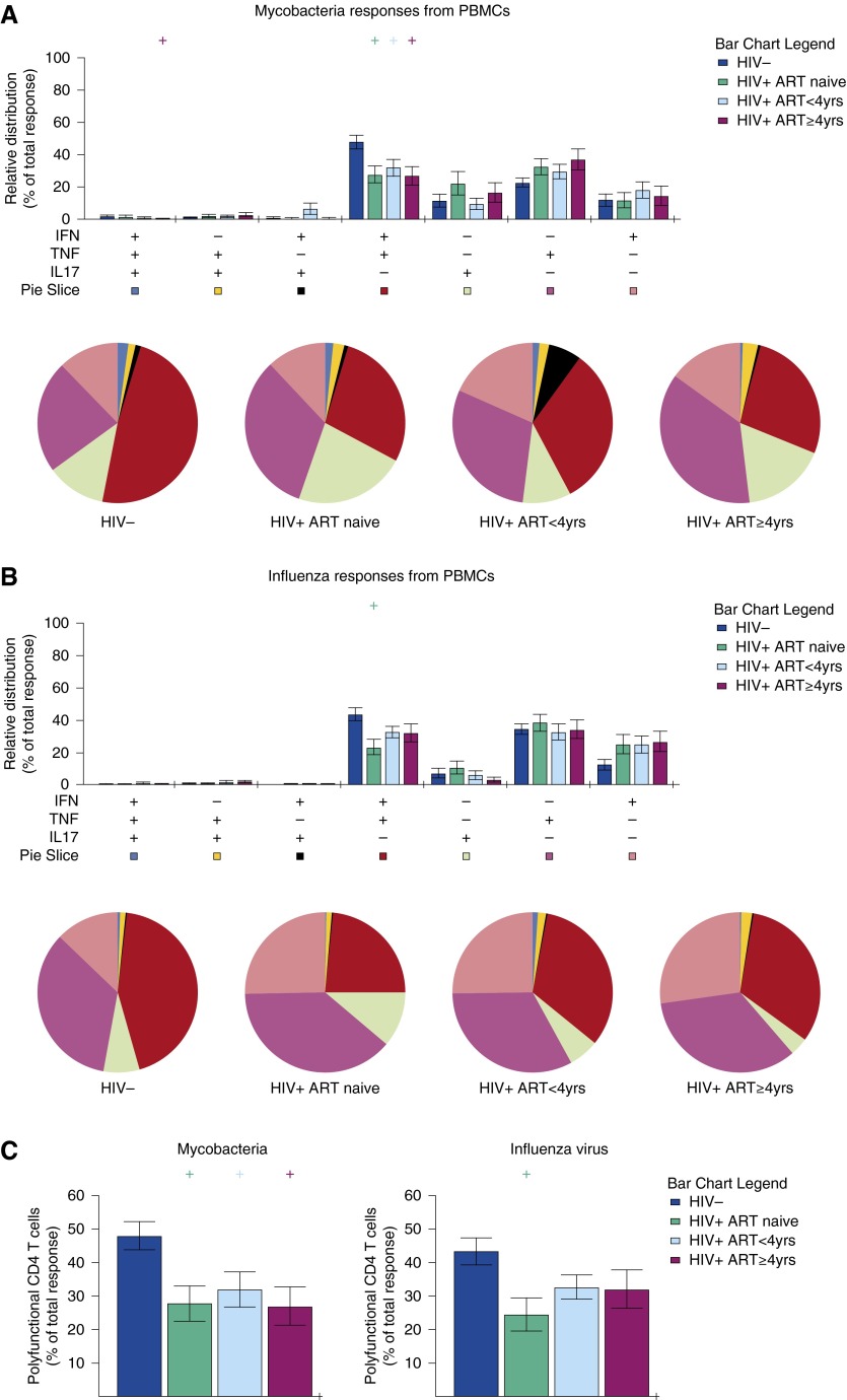
Measurements and main results: HIV-infected adults on ART exhibited lower plasma HIV viral load and higher blood CD4(+) T-cell count than ART-naive adults. AM proteolysis and total mycobacteria-specific Th1 CD4(+) T-cell responses in individuals on ART for greater than or equal to 4 years were similar to HIV-uninfected control subjects but those on ART for less than 4 years had impaired responses. Total influenza-specific alveolar Th1 CD4(+) T-cell responses were intact in all individuals receiving ART. In contrast, BAL and blood mycobacteria-specific polyfunctional CD4(+) T-cell responses were impaired in adults on ART irrespective of duration.

Conclusions: AM and mycobacteria-specific alveolar CD4(+) T-cell responses in HIV-infected adults on ART for less than 4 years are impaired and may partly explain the high risk of TB in HIV-infected individuals on ART. Strategies to augment ART to improve lung immune cell function and reduce the high incidence of TB in HIV-infected adults who initiate ART should be investigated.

Keywords: ART; HIV; Mycobacterium tuberculosis; T cells; macrophages.


Figures

Figure 1.
Figure 1.
Impaired alveolar macrophage proteolysis function in HIV-infected adults on antiretroviral therapy (ART) for less than 4 years. Adherent alveolar macrophages were incubated with DQ-BSA reporter beads for 10 minutes or 240 minutes to measure phagosomal bulk proteolysis. The cells were acquired on a flow cytometer and the activity index was determined. Data were log transformed. Gray bars represent geometric mean and black horizontal lines represent confidence intervals. Data were analyzed using one-way analysis of variance and Student t test (HIV+ ART naive, n = 18; HIV+ ART < 4 yr, n = 15; HIV+ ART ≥ 4 yr, n = 16; HIV, n = 19).
Figure 2.
Figure 2.
Representative flow cytometry dot plot from an HIV-uninfected adult showing multiple subsets of antigen-specific CD4+ T cells in bronchoalveolar lavage (BAL). Nonadherent BAL cells and peripheral blood mononuclear cells were stimulated overnight with purified protein derivative or influenza vaccine for 18 hours and CD4+ T-cell responses were measured by intracellular cytokine staining. The dot plots were obtained by gating on singlets, lymphocytes, live cells, CD3+ cells, CD4+ cells, and combination of three cytokines. They show the frequency (percentage) of IFN-γ–, tumor necrosis factor (TNF)-α–, and/or IL-17–producing CD4+ T cells in BAL, in an unstimulated negative control and cells stimulated with PMA/ionomycin (positive control), mycobacteria, and influenza virus antigens. PMA = phorbol myristate acetate.
Figure 3.
Figure 3.
Impaired total mycobacteria-specific alveolar Th1 CD4+ T-cell responses in HIV-infected adults on antiretroviral therapy (ART) for less than 4 years. Nonadherent bronchoalveoalar lavage (BAL) cells were stimulated overnight with purified protein derivative and influenza vaccine. Cells were analyzed by flow cytometry for IFN-γ and tumor necrosis factor production by the CD4+ T-cell population. (A and C) Mycobacterium-specific responses in BAL and peripheral blood mononuclear cells (PBMCs). (B and D) Influenza-specific responses in BAL and PBMCs. The data were log transformed and are shown as the total frequency of antigen-specific Th1 IFN-γ– and tumor necrosis factor–producing CD4+ T cells. The horizontal bars represent geometric mean and confidence interval. Data were analyzed using one-way analysis of variance and Student t test (HIV, n = 32; HIV+ ART naive, n = 25; HIV+ ART < 4 yr, n = 28; HIV+ ART ≥ 4 yr, n = 22).
Figure 4.
Figure 4.
Impaired polyfunctional mycobacteria-specific alveolar CD4+ T cells in HIV-infected adults on antiretroviral therapy (ART). Nonadherent bronchoalveoalar lavage (BAL) cells were stimulated overnight with (A) purified protein derivative and (B) influenza vaccine. Cells were analyzed by flow cytometry for IFN-γ, tumor necrosis factor (TNF), and IL-17 production by the CD4+ T-cell population. Data are shown as relative distribution of IFN-γ–, TNF–, and IL-17–producing CD4+ T-cell subsets within the total response. Bar charts represent the mean and SEM of the contribution of the indicated subset (x axis) toward the total antigen-specific CD4+ T-cell response against the indicated participant groups (color coded as shown). P values were computed using Student t test (denoted as “+“ if P < 0.05), comparing each group against HIV-uninfected adults. Each pie chart represents the mean distribution across subjects of different antigen-specific cytokine-producing CD4+ T-cell subsets (color coded as shown) within the total response in a particular group. (C) Relative distribution of polyfunctional IFN-γ– and TNF-producing CD4+ T cells within the total response. (HIV, n = 32; HIV+ ART naive, n = 25; HIV+ ART < 4 yr, n = 24; HIV+ ART ≥ 4 yr, n = 15).
Figure 5.
Figure 5.
Impaired polyfunctional mycobacteria-specific blood CD4+ T cells in HIV-infected adults on antiretroviral therapy (ART). Peripheral blood mononuclear cells (PBMCs) were stimulated overnight with (A) purified protein derivative and (B) influenza vaccine. Cells were analyzed by flow cytometry for IFN-γ, tumor necrosis factor (TNF), and IL-17 production by the CD4+ T-cell population. Data are shown as relative distribution of IFN-γ–, TNF–, and IL-17–producing CD4+ T-cell subsets within the total response. Bar charts represent the mean and SEM of the contribution of the indicated subset (x axis) toward the total antigen-specific CD4+ T-cell response against the indicated participant groups (color coded as shown). P values were computed using Student t test (denoted as “+ “ if P < 0.05), comparing each group against HIV-uninfected adults. Each pie chart represents the mean distribution across subjects of different antigen-specific cytokine-producing CD4+ T-cell subsets (color coded as shown) within the total response in a particular group. (C) Relative distribution of polyfunctional IFN-γ– and TNF-producing CD4+ T cells within the total response. (HIV, n = 32; HIV+ ART naive, n = 25; HIV+ ART < 4 yr, n = 28; HIV+ ART ≥ 4 yr, n = 22).

References

    1. Harries AD, Zachariah R, Corbett EL, Lawn SD, Santos-Filho ET, Chimzizi R, Harrington M, Maher D, Williams BG, De Cock KM. The HIV-associated tuberculosis epidemic—when will we act? Lancet. 2010;375:1906–1919. - PubMed
    1. Sonnenberg P, Glynn JR, Fielding K, Murray J, Godfrey-Faussett P, Shearer S. How soon after infection with HIV does the risk of tuberculosis start to increase? A retrospective cohort study in South African gold miners. J Infect Dis. 2005;191:150–158. - PubMed
    1. Houben RM, Crampin AC, Ndhlovu R, Sonnenberg P, Godfrey-Faussett P, Haas WH, Engelmann G, Lombard CJ, Wilkinson D, Bruchfeld J, et al. Human immunodeficiency virus associated tuberculosis more often due to recent infection than reactivation of latent infection. Int J Tuberc Lung Dis. 2011;15:24–31. - PubMed
    1. Kalsdorf B, Scriba TJ, Wood K, Day CL, Dheda K, Dawson R, Hanekom WA, Lange C, Wilkinson RJ. HIV-1 infection impairs the bronchoalveolar T-cell response to mycobacteria. Am J Respir Crit Care Med. 2009;180:1262–1270. - PMC - PubMed
    1. Jambo KC, Sepako E, Fullerton DG, Mzinza D, Glennie S, Wright AK, Heyderman RS, Gordon SB. Bronchoalveolar CD4+ T cell responses to respiratory antigens are impaired in HIV-infected adults. Thorax. 2011;66:375–382. - PMC - PubMed

Publication types

MeSH terms

Substances