. 2014 Sep 16:5:4816.
doi: 10.1038/ncomms5816.

Conserved and host-specific features of influenza virion architecture

Affiliations

Conserved and host-specific features of influenza virion architecture

Edward C Hutchinson et al. Nat Commun. .

Erratum in

Abstract

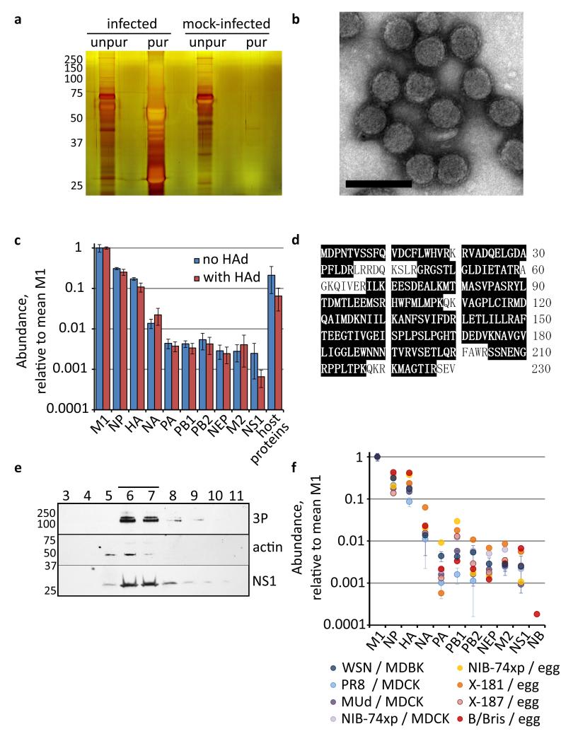
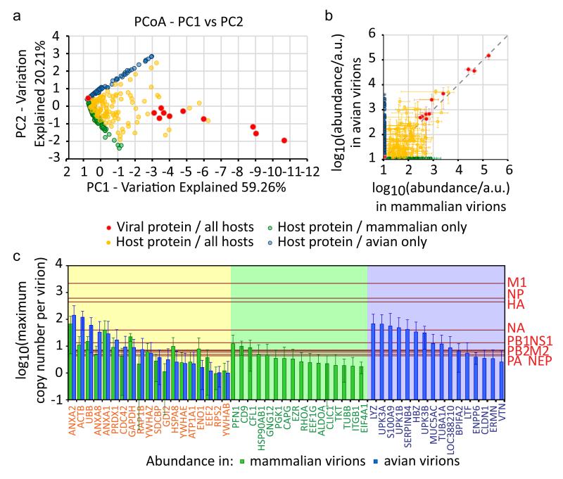
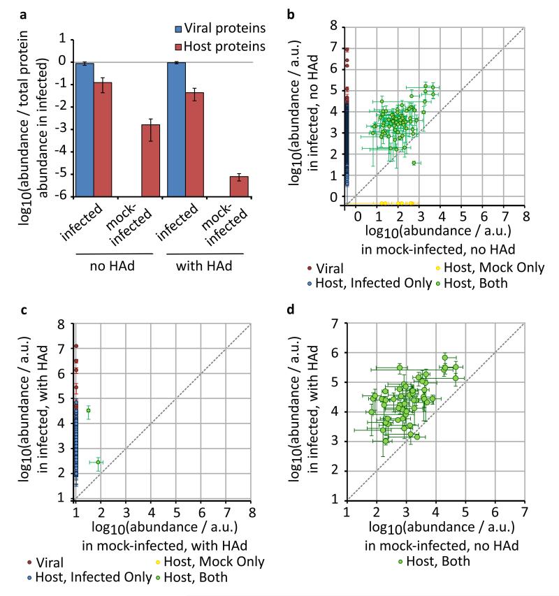
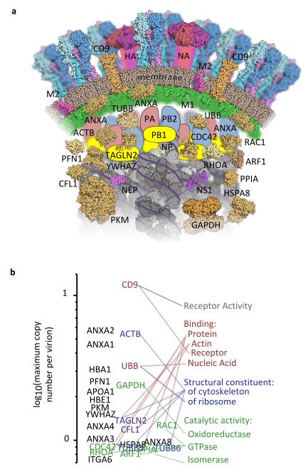
Viruses use virions to spread between hosts, and virion composition is therefore the primary determinant of viral transmissibility and immunogenicity. However, the virions of many viruses are complex and pleomorphic, making them difficult to analyse in detail. Here we address this by identifying and quantifying virion proteins with mass spectrometry, producing a complete and quantified model of the hundreds of host-encoded and viral proteins that make up the pleomorphic virions of influenza viruses. We show that a conserved influenza virion architecture is maintained across diverse combinations of virus and host. This 'core' architecture, which includes substantial quantities of host proteins as well as the viral protein NS1, is elaborated with abundant host-dependent features. As a result, influenza virions produced by mammalian and avian hosts have distinct protein compositions. Finally, we note that influenza virions share an underlying protein composition with exosomes, suggesting that influenza virions form by subverting microvesicle production.


Figures

Figure 1
Figure 1. The abundance of viral proteins in influenza virions
Bovine epithelial (MDBK) cells were infected with influenza A/WSN/33 virus (WSN) or mock-infected, and media harvested after 48 h. (a) Samples of media were separated by SDS-PAGE either before (diluted 1/1000) or after purification by gradient ultracentrifugation, and silver-stained. (b) Purified influenza A/WSN/33 virions, negatively-stained and visualised by EM; the scale bar is 200 nm. (c) Abundance of viral proteins in purified virions, calculated by SINQ and normalized by the mean level of M1. The total abundance of all host proteins detected is also shown. The mean and s.d. is shown of 3 separate experiments, performed either with or without a haemadsorption / elution step (HAd). (d) The NS1 sequence of WSN shaded to show peptides identified by LC-MS/MS (from 3 separate experiments, purified without HAd; 87 % coverage). (e) Virions were layered onto a 30 – 60 % sucrose gradient and subjected to ultracentrifugation. The gradient was then harvested in 15 fractions from the top; the fractions shown were analysed by 14% SDS-PAGE and Western blotting for the indicated proteins. The position of molecular weight markers is indicated in kDa; concentrated virions were visible as a band in the gradient between fractions 6 and 7 (line). (f) Abundance of viral proteins in virions of 6 different influenza A viruses and an influenza B virus, grown in a variety of hosts (bovine epithelial (MDBK) cells, canine epithelial (MDCK) cells, and embryonated chicken eggs; see Table 1 for details). For WSN the mean and s.d. of 3 separate experiments is shown and for PR8 and MUd the mean and range of 2 separate experiments.
Figure 2
Figure 2. The abundance of host proteins in influenza virions
Protein abundance in purified virions was determined by SINQ. A threshold was set at one tenth the abundance of the least abundant polymerase subunit, and any protein of lower abundance was assigned this value. Proteins were matched to their human orthologs to allow comparisons between virions from different host species. (a) Principal co-ordinates analysis of log10(protein abundance/a.u.) for 548 protein orthologs (goodness of fit = 0.795). (b) Log10(abundance/a.u.) of proteins in virions from mammalian cells and in virions from avian cells; mean and s.d. of four combinations of viruses and mammalian hosts, and four combinations of viruses and avian hosts (Table 1). (c) The core and host-dependent features of influenza virions. The maximum copy number of proteins in virions is shown, calculated by assuming that an average virion incorporates no more than eight viral polymerases. Viral proteins are shown as red lines, and host proteins with a mean copy number greater than one are shown as bars. Host proteins are only shown if detected in all four combinations of virus and host with both mammalian hosts (green bars) and avian hosts (blue bars); the mean and s.d. is indicated. The core architecture of a spherical influenza virion consists of the viral proteins (red lines) and those host proteins found in all hosts (yellow background). Additional host proteins are found only in virions from mammalian hosts (green background) or from avian hosts (blue background).
Figure 3
Figure 3. Comparison of proteins shed by infected and uninfected cells
To compare shedding from infected and mock-infected cells, gradient ultracentrifugation was used to purify virion-sized material from the growth media of bovine epithelial (MDBK) cells 48 h after either infection with WSN or mock-infection. Purifications were performed with or without a prior haemadsorption / elution step (HAd), and protein standards were added after purification to allow comparison of samples with different total protein concentrations. Purified proteins were quantified by SINQ. (a) Total abundance of purified viral and host proteins, relative to the mean total protein abundance in the infected samples (mean and s.d. of three separate experiments, not including protein standards). (bd) The abundance of individual proteins shed by infected and mock-infected cells, and detected in at least two of three separate experiments (mean and s.d. if N=3, mean and range if N=2; not including protein standards). Proteins not detected in one condition (infected or mock-infected) were assigned the lowest abundance of any protein detected in that condition. Panels show the abundance of proteins (b) purified without HAd, (c) purified with HAd, and (d) common to the mock-infected sample without HAd and to the infected sample with HAd.
Figure 4
Figure 4. The architecture of an influenza virion
Proteins in virions produced by WSN-infected bovine epithelial (MDBK) cells, present at more than one tenth the abundance of the viral polymerase and found in material purified both with and without HAd in at least two separate experiments. (a) Schematic cross-section of an influenza virion, showing proteins whose localisation in the virion is known or can be inferred from other studies. Host proteins and membrane are brown and viral proteins are brightly coloured. (b) Host proteins, ranked by maximum copy number in virions (calculated as in Fig. 2) and linked to their Gene Ontology molecular function terms.

References

    1. Harris A, et al. Influenza virus pleiomorphy characterized by cryoelectron tomography. Proc Natl Acad Sci U S A. 2006;103:19123–19127. doi:10.1073/pnas.0607614103. - PMC - PubMed
    1. Noda T, et al. Architecture of ribonucleoprotein complexes in influenza A virus particles. Nature. 2006;439:490–492. doi:10.1038/nature04378. - PubMed
    1. Wasilewski S, Calder LJ, Grant T, Rosenthal PB. Distribution of surface glycoproteins on influenza A virus determined by electron cryotomography. Vaccine. 2012;30:7368–7373. doi:10.1016/j.vaccine.2012.09.082. - PMC - PubMed
    1. Vijayakrishnan S, et al. Cryotomography of budding influenza A virus reveals filaments with diverse morphologies that mostly do not bear a genome at their distal end. PLoS Pathog. 2013;9:e1003413. doi:10.1371/journal.ppat.1003413. - PMC - PubMed
    1. Nermut MV. Further investigation on the fine structure of influenza virus. J Gen Virol. 1972;17:317–331. - PubMed

Publication types

MeSH terms

Substances