. 2014 Sep 16;5(5):e01690-14.
doi: 10.1128/mBio.01690-14.

Streptolysin O and NAD-glycohydrolase prevent phagolysosome acidification and promote group A Streptococcus survival in macrophages

Affiliations

Streptolysin O and NAD-glycohydrolase prevent phagolysosome acidification and promote group A Streptococcus survival in macrophages

Benedicte Bastiat-Sempe et al. mBio. .

Abstract

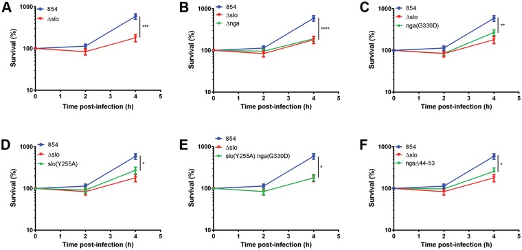
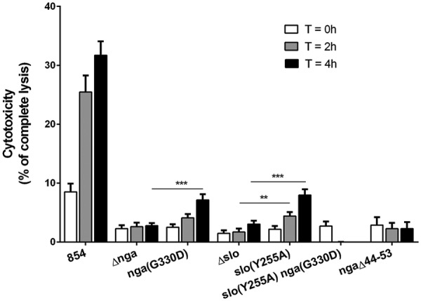
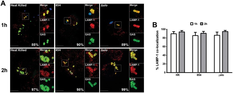
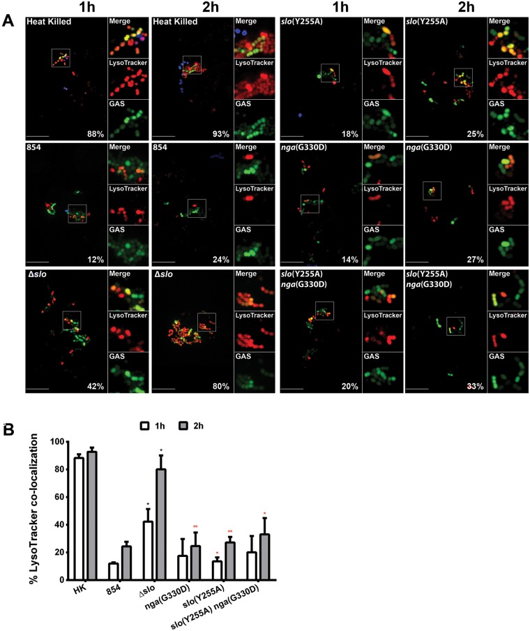
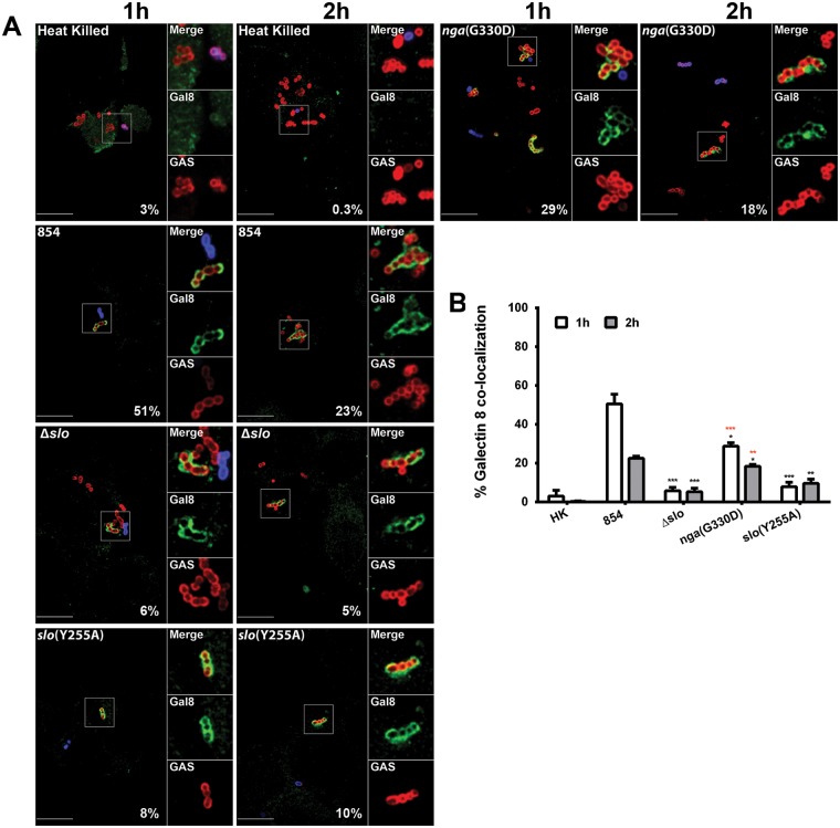
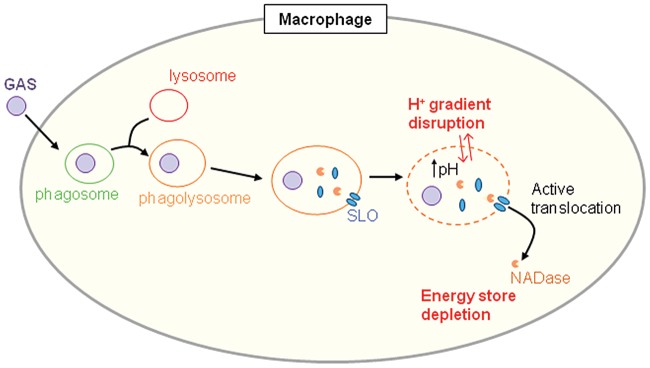
Group A Streptococcus (GAS, Streptococcus pyogenes) is an ongoing threat to human health as the agent of streptococcal pharyngitis, skin and soft tissue infections, and life-threatening conditions such as necrotizing fasciitis and streptococcal toxic shock syndrome. In animal models of infection, macrophages have been shown to contribute to host defense against GAS infection. However, as GAS can resist killing by macrophages in vitro and induce macrophage cell death, it has been suggested that GAS intracellular survival in macrophages may enable persistent infection. Using isogenic mutants, we now show that the GAS pore-forming toxin streptolysin O (SLO) and its cotoxin NAD-glycohydrolase (NADase) mediate GAS intracellular survival and cytotoxicity for macrophages. Unexpectedly, the two toxins did not inhibit fusion of GAS-containing phagosomes with lysosomes but rather prevented phagolysosome acidification. SLO served two essential functions, poration of the phagolysosomal membrane and translocation of NADase into the macrophage cytosol, both of which were necessary for maximal GAS intracellular survival. Whereas NADase delivery to epithelial cells is mediated by SLO secreted from GAS bound to the cell surface, in macrophages, the source of SLO and NADase is GAS contained within phagolysosomes. We found that transfer of NADase from the phagolysosome to the macrophage cytosol occurs not by simple diffusion through SLO pores but rather by a specific translocation mechanism that requires the N-terminal translocation domain of NADase. These results illuminate the mechanisms through which SLO and NADase enable GAS to defeat macrophage-mediated killing and provide new insight into the virulence of a major human pathogen.

Importance: Macrophages constitute an important element of the innate immune response to mucosal pathogens. They ingest and kill microbes by phagocytosis and secrete inflammatory cytokines to recruit and activate other effector cells. Group A Streptococcus (GAS, Streptococcus pyogenes), an important cause of pharyngitis and invasive infections, has been shown to resist killing by macrophages. We find that GAS resistance to macrophage killing depends on the GAS pore-forming toxin streptolysin O (SLO) and its cotoxin NAD-glycohydrolase (NADase). GAS bacteria are internalized by macrophage phagocytosis but resist killing by secreting SLO, which damages the phagolysosome membrane, prevents phagolysosome acidification, and translocates NADase from the phagolysosome into the macrophage cytosol. NADase augments SLO-mediated cytotoxicity by depleting cellular energy stores. These findings may explain the nearly universal production of SLO by GAS clinical isolates and the association of NADase with the global spread of a GAS clone implicated in invasive infections.


Figures

FIG 1
FIG 1
SLO and NADase are required for maximal survival of GAS in macrophages. Values are the mean numbers of CFU recovered immediately after 90 min of exposure to GAS (time zero) and 2 and 4 h later, expressed as a percentage of the value at time zero. Data for wild-type GAS strain 854 and the SLO-deficient Δslo mutant strain are shown in panel A and for clarity in panels B through F in comparison to those of individual mutant strains. Data represent the mean values ± the standard errors from at least four experiments performed in triplicate. *, P < 0.05; **, P < 0.01; ***, P < 0.001; ****, P < 0.0001.
FIG 2
FIG 2
SLO and NADase mediate GAS cytotoxicity for macrophages. Cell culture supernatants were collected to measure the LDH released by macrophages at 0, 2, and 4 h after infection. Cytotoxicity is expressed as a percentage of the amount of LDH released by uninfected macrophages lysed with 10% Triton X-100. Shown are the mean values ± the standard errors of four independent experiments performed in triplicate. Values for all of the mutant strains were significantly different from those for strain 854 at the corresponding time points. Asterisks indicate other comparisons with statistically significant differences. **, P < 0.01; ***, P < 0.001.
FIG 3
FIG 3
SLO and NADase do not prevent lysosome fusion to the GAS-containing phagosome. (A) Confocal microscopy images showing colocalization of LAMP-1 (Alexa 568, red) and heat-killed GAS strain 854 (HK), live 854, or the Δslo mutant (Alexa 488, green). Extracellular bacteria were stained both green and blue (Alexa 660). Representative images from one experiment are shown. Scale bars = 10 µm. The percentage of intracellular bacteria of each strain colocalized with LAMP-1 is indicated. (B) Percentage of intracellular GAS associated with LAMP-1 after 1 h (white bars) or 2 h (gray bars) of infection. Mean values ± the standard errors of at least three independent experiments are shown.
FIG 4
FIG 4
SLO impairs acidification of GAS-containing phagolysosomes. (A) Confocal microscopy images demonstrating the association between the acidotropic probe LysoTracker Red DND-99 (red) and GFP-expressing GAS (green) at 1 and 2 h of infection. Extracellular GAS bacteria are also stained blue (Alexa 660). Representative images from one experiment are shown. Scale bars = 10 µm. The percentage of intracellular bacteria of each strain that colocalized with LysoTracker is shown. (B) Percentage of heat-killed (HK) or live GAS colocalized with LysoTracker at 1 h (white bars) or 2 h (gray bars) of infection. Mean values ± the standard errors of at least three independent experiments are shown. Black asterisks indicate values that differ significantly from that for strain 854; red asterisks indicates values that differ significantly from that for the Δslo mutant. *, P < 0.05; **, P < 0.01.
FIG 5
FIG 5
SLO mediates membrane damage to GAS-containing phagolysosomes. (A) Confocal microscopy images of the association between GAS (Alexa 568, red) and galectin 8 (Alexa 488, green) during the infection of macrophages. Extracellular GAS bacteria are both red and blue (Alexa 568 and 660, respectively). Images representative of one experiment are shown. Scale bars = 10 µm. The percentage of intracellular bacteria of each strain associated with galectin 8 is indicated. (B) Percentage of colocalization of GAS and galectin 8 after 1 h (white bars) or 2 h (gray bars) of infection. Mean values ± the standard errors of at least three independent experiments are shown. Black asterisks indicate values that differ significantly from that of strain 854, and red asterisks indicate values that differ significantly from that of the Δslo mutant strain. *, P < 0.05; **, P < 0.01; ***, P < 0.001. HK, heat killed.
FIG 6
FIG 6
Model of GAS survival in macrophages. GAS is internalized by phagocytosis. The GAS-containing phagosome fuses with lysosomes to form a phagolysosome. SLO and NADase are secreted by GAS into this compartment. SLO damages the phagolysosome membrane, preventing phagolysosome acidification. NADase is translocated by SLO from the phagolysosome into the macrophage cytosol, where it depletes NAD+ and ATP, thereby inhibiting cellular repair of SLO-mediated membrane damage. The combined action of SLO and NADase interferes with phagolysosomal killing and promotes GAS intracellular survival.

References

    1. Cunningham MW. 2000. Pathogenesis of group A streptococcal infections. Clin. Microbiol. Rev. 13:470–511. 10.1128/CMR.13.3.470-511.2000 - DOI - PMC - PubMed
    1. Osterlund A, Popa R, Nikkilä T, Scheynius A, Engstrand L. 1997. Intracellular reservoir of Streptococcus pyogenes in vivo: a possible explanation for recurrent pharyngotonsillitis. Laryngoscope 107:640–647. 10.1097/00005537-199705000-00016 - DOI - PubMed
    1. Spinaci C, Magi G, Varaldo PE, Facinelli B. 2006. Persistence of erythromycin-resistant group A streptococci in cultured respiratory cells. Pediatr. Infect. Dis. J. 25:880–883. 10.1097/01.inf.0000238136.63851.4a - DOI - PubMed
    1. Rohde M, Chhatwal GS. 2013. Adherence and invasion of streptococci to eukaryotic cells and their role in disease pathogenesis. Curr. Top. Microbiol. Immunol. 368:83–110. 10.1007/82_2012_281 - DOI - PubMed
    1. Medina E, Rohde M, Chhatwal GS. 2003. Intracellular survival of Streptococcus pyogenes in polymorphonuclear cells results in increased bacterial virulence. Infect. Immun. 71:5376–5380. 10.1128/IAI.71.9.5376-5380.2003 - DOI - PMC - PubMed

Publication types