. 2014;35(3):415-26.
doi: 10.3233/NRE-141132.

Differential patterns of cortical reorganization following constraint-induced movement therapy during early and late period after stroke: A preliminary study

Affiliations

Differential patterns of cortical reorganization following constraint-induced movement therapy during early and late period after stroke: A preliminary study

Lumy Sawaki et al. NeuroRehabilitation. 2014.

Abstract

Objective: Constraint-induced movement therapy (CIMT) has been shown to improve upper extremity voluntary movement and change cortical movement representation after stroke. Direct comparison of the differential degree of cortical reorganization according to chronicity in stroke subjects receiving CIMT has not been performed and was the purpose of this study. We hypothesized that a higher degree of cortical reorganization would occur in the early (less than 9 months post-stroke) compared to the late group (more than 12 months post-stroke).

Methods: 17 early and 9 late subjects were enrolled. Each subject was evaluated using transcranial magnetic stimulation (TMS) and the Wolf Motor Function Test (WMFT) and received CIMT for 2 weeks.

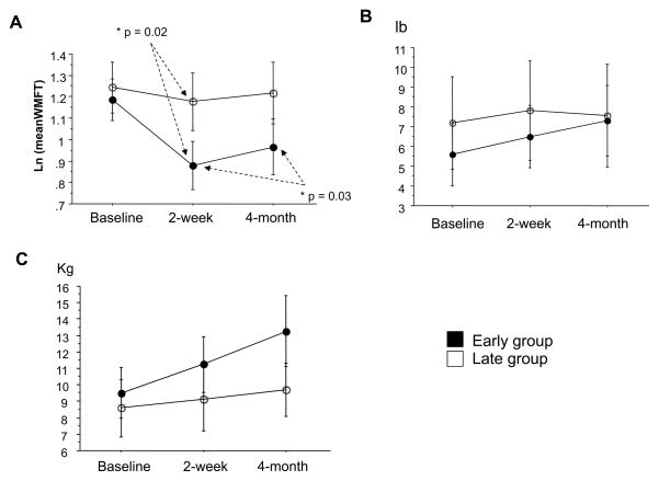
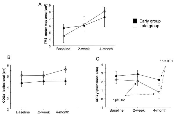
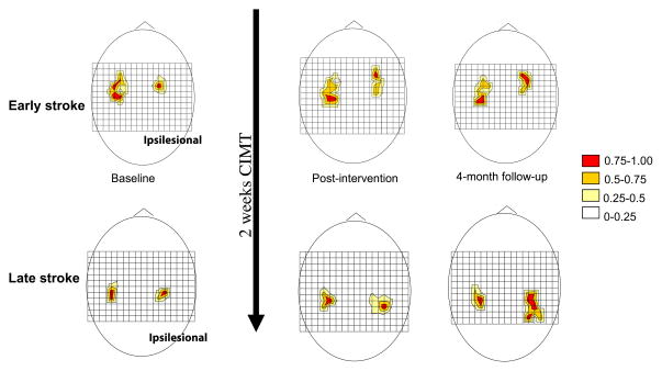
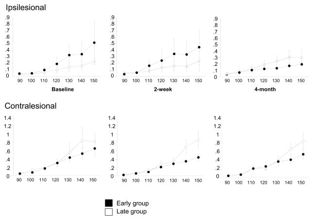
Results: The early group showed greater improvement in WMFT compared with the late group. TMS motor maps showed persistent enlargement in both groups but the late group trended toward more enlargement. The map shifted posteriorly in the late stroke group. The main limitation was the small number of TMS measures that could be acquired due to high motor thresholds, particularly in the late group.

Conclusion: CIMT appears to lead to greater improvement in motor function in the early phase after stroke. Greater cortical reorganization in map size and position occurred in the late group in comparison.

Significance: The contrast between larger functional gains in the early group vs larger map changes in the late group may indicate that mechanisms of recovery change over the several months following stroke or that map changes are a time-dependent epiphenomenon.

Keywords: Plasticity; motor; recovery; transcranial magnetic stimulation; upper extremity.


Figures

Figure 1
Figure 1
Effect of CIMT on Wolf Motor Function Test (WMFT) collected at baseline, at 2 weeks (ie, completion of intervention), and at 4-month follow-up. A: Log mean of time-based evaluations (scores indicate time to complete tasks; smaller scores indicate greater improvement). B: Mean of force-based measure (lift weight lb; higher scores indicate more weight lifted). C: Mean of force-based measure (grip strength kg; higher scores indicate more grip strength). Note that both early (solid dots) and late (open dots) stroke groups showed improvement in all performance measures at 2 weeks and at 4-month follow-up. Time-based measures in early group showed significant improvement compared with the late group immediately after CIMT (P = .024). Data are expressed as mean ± SE.
Figure 2
Figure 2
Longitudinal changes in TMS motor map volume (A), COG x (B) and COG y (C) on the ipsilesional hemisphere. Note that both early (solid dots) and late (open dots) stroke groups exhibit increased map volume after 2 weeks and at 4-month follow-up. There is, however, a significantly further posterior shift of COG y from its position at 2 weeks to its position at 4-month follow-up for the late group as compared with that of the early group (Figure 2C, P = .01). Additionally, there is a significant difference of COG y between the early and late groups at 4-month follow-up (Figure 2C, P = .02), suggesting a differential pattern of reorganization between the groups. Data are expressed as mean ± SE.
Figure 3
Figure 3
Longitudinal changes in TMS motor map volume of 2 representative subjects. The grid size is 1 cm, and (0,0) is Cz in the 10–20 EEG system. Motor responses at each scalp position are color-coded by MEP size (relative to the maximal response). Increased TMS motor map volume of ipsilesional hemisphere was observed in both an early subject (top diagrams) and a late subject (bottom diagrams) over a 4-month period. The 4-month follow-up for the late subject showed a significantly posterior shift of TMS motor map.
Figure 4
Figure 4
Longitudinal changes in recruitment curves for ipsilesional (upper graphs) and contralesional (bottom graphs). Note that while there is no significant difference between the early (solid dots) and late (open dots) groups at any time, there is a reversed pattern in the ipsilesional and contralesional hemispheres particularly in the late group. That is to say that, the ipsilesional hemisphere in the late group appears to be less excitable in response to TMS at baseline and the contralesional hemisphere exhibits higher corticomotor excitability. This pattern is, however, lost at 4-month follow up. Data are expressed as mean ± SE.

References

    1. Alberts JL, Butler AJ, Wolf SL. The effects of constraint-induced therapy on precision grip: a preliminary study. Neurorehabil Neural Repair. 2004;18:250–8. - PMC - PubMed
    1. Barbay S, Plautz EJ, Friel KM, Frost SB, Dancause N, Stowe AM, et al. Behavioral and neurophysiological effects of delayed training following a small ischemic infarct in primary motor cortex of squirrel monkeys. Exp Brain Res. 2006;169:106–16. - PMC - PubMed
    1. Bastings EP, Greenberg JP, Good DC. Hand motor recovery after stroke: a transcranial magnetic stimulation mapping study of motor output areas and their relation to functional status. Neurorehabil Neural Repair. 2002;16:275–82. - PubMed
    1. Blanton S, Wolf SL. An application of upper-extremity constraint-induced movement therapy in a patient with subacute stroke. Phys Ther. 1999;79:847–53. - PubMed
    1. Bonifer NM, Anderson KM, Arciniegas DB. Constraint-induced therapy for moderate chronic upper extremity impairment after stroke. Brain Inj. 2005;19:323–30. - PubMed

Publication types