. 2015 May;8(3):505-15.
doi: 10.1038/mi.2014.84. Epub 2014 Sep 17.

Dual-specificity phosphatase 6 regulates CD4+ T-cell functions and restrains spontaneous colitis in IL-10-deficient mice

Affiliations

Dual-specificity phosphatase 6 regulates CD4+ T-cell functions and restrains spontaneous colitis in IL-10-deficient mice

S Bertin et al. Mucosal Immunol. 2015 May.

Abstract

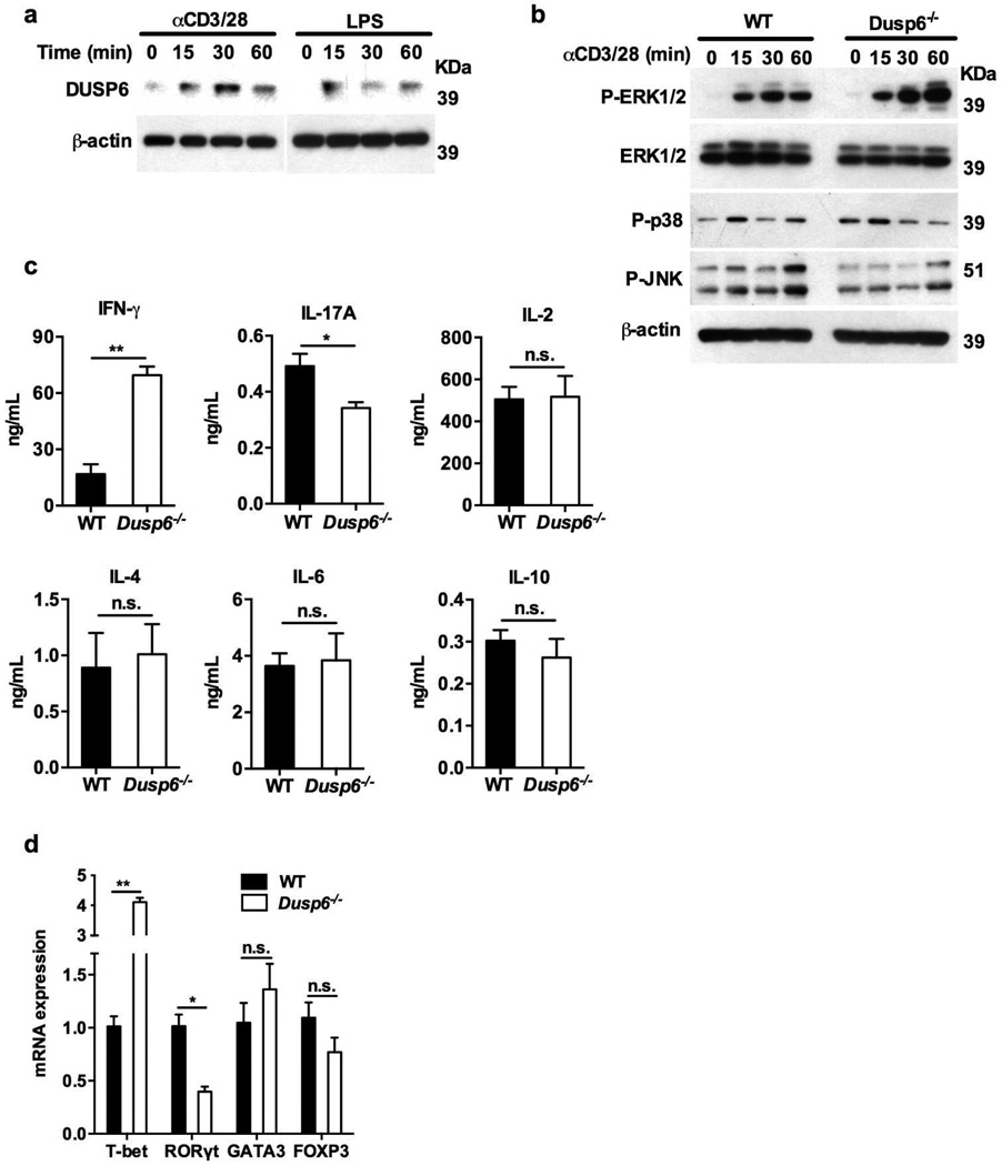
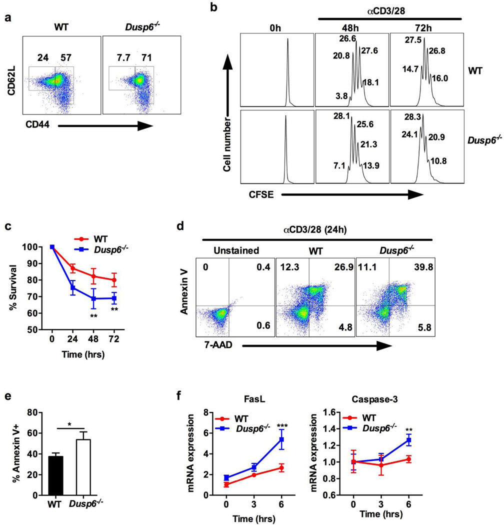
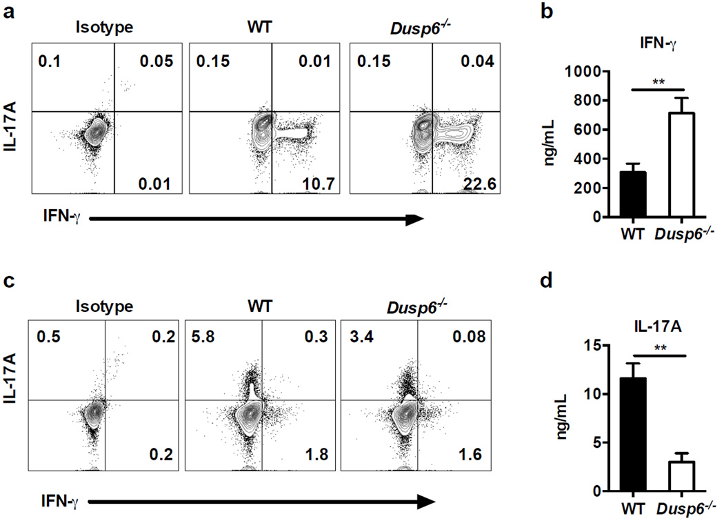
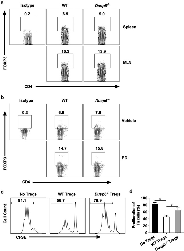
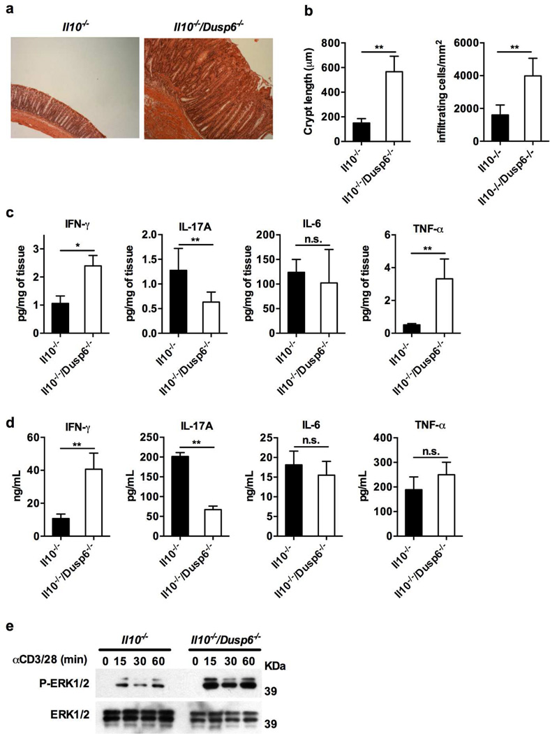
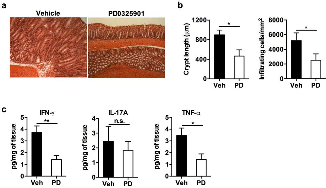
Mitogen-activated protein kinase (MAPK) phosphatases are dual-specificity phosphatases (DUSPs) that dephosphorylate phosphothreonine and phosphotyrosine residues within MAPKs. DUSP6 preferentially dephosphorylates extracellular signal-regulated kinases 1 and 2 (ERK1/2) rendering them inactive. Here, we study the role of DUSP6 in CD4(+) T-cell function, differentiation, and inflammatory profile in the colon. Upon T-cell receptor (TCR) stimulation, DUSP6 knockout (Dusp6(-/-)) CD4(+) T cells showed increased ERK1/2 activation, proliferation, T helper 1 differentiation, and interferon-γ production, as well as a marked decrease in survival, interleukin- 17A (IL-17A) secretion, and regulatory T-cell function. To analyze the role of DUSP6 in vivo, we employed the Il10(-/-) model of colitis and generated Il10(-/-)/Dusp6(-/-) double-knockout mice. Il10(-/-)/Dusp6(-/-) mice suffered from accelerated and exacerbated spontaneous colitis, which was prevented by ERK1/2 inhibition. ERK1/2 inhibition also augmented regulatory T-cell differentiation in vitro and in vivo in both C57Bl/6 and Dusp6(-/-) mice. In summary, DUSP6 regulates CD4(+) T-cell activation and differentiation by inhibiting the TCR-dependent ERK1/2 activation. DUSP6 might therefore be a potential intervention target for limiting aberrant T-cell responses in T-cell-mediated diseases, such as inflammatory bowel disease.


Conflict of interest statement

Conflict of interest

The authors declare no conflict of interest.

Figures

Figure 1
Figure 1. Increased ERK1/2 activation and IFN-γ production in Dusp6−/−CD4+ T cells
(a) Immunoblot analysis of DUSP6 expression in CD4+ T cells from WT mice upon activation of either TLR4 (100 µg LPS) or T cell receptor (αCD3/28 antibodies). (b) Immunoblot analysis of phosphorylated levels of MAPKs in CD4+ T cells stimulated with anti-CD3/28 antibodies for the indicated time-points. Data are representative of three independent experiments (a,b). (c) Cytokine levels in culture supernatants of MLN-derived CD4+ T cells isolated from WT and Dusp6−/− mice and stimulated with anti-CD3/28 antibodies for 24 hours (n≥9 mice per group). (d) qPCR analysis of the expression of different transcription factors in CD4+ T cells from WT and Dusp6−/− mice stimulated with anti-CD3/28 antibodies (n≥5 mice per group). The mRNA fold induction in Dusp6−/− CD4+ T cells was normalized according to the mRNA expression in WT T cells. GAPDH was used as internal control. Error bars represent standard deviation Data represent results from either three (a–c) or two (d) independent experiments. *P<0.05, **P<0.01.
Figure 2
Figure 2. DUSP6 deficiency results in increased proliferation and activation-induced cell death
(a) Flow cytometry analysis of the naïve (CD44lowCD62Lhigh) and activated (CD44high) CD4+ T cell populations in spleens of WT and Dusp6−/− mice. (b) Proliferation assay by flow cytometry analysis of the fluorescence intensity of CFSE-labeled naïve T cells from WT and Dusp6−/− mice after 48 and 72 hours of TCR stimulation with anti-CD3/28 antibodies. Data are representative of two independent experiments (a,b). (c) Percent of viable CD4+ T cells measured by microscopic evaluation using the trypan blue exclusion method in WT and Dusp6−/− T cells stimulated with anti-CD3/CD28 antibodies. Data represent pooled results from three independent experiments. **P<0.01 (d) Flow cytometry analysis of early apoptotic (Annexin V+, 7-AAD), late stage apoptotic (Annexin V+, 7-AAD+), and necrotic (Annexin V, 7-AAD+) CD4+ T cells after 24 hours of stimulation with anti-CD3/28 antibodies. Data are representative of three independent experiments (n=3 mice per group). (e) Statistical analysis of the percentage of total Annexin V+ cells shown in d) analysis. Data represents pooled results from three independent experiments (n=3 mice per group). (f) qPCR analysis of pro-apoptotic genes in WT and Dusp6−/− CD4+ T cells stimulated with anti-CD3/28 antibodies. Data represent pooled results from two experiments (n=3 mice/group). Error bars representmean ± standard deviation. **P<0.01, ***P<0.001
Figure 3
Figure 3. DUSP6 deficiency facilitates Th1 differentiation in vitro
(a) and (c) Flow cytometry analysis of intracellular cytokines in naïve CD4+ T cells from WT and Dusp6−/− mice cultured in Th1 (a) or Th17 (c) polarizing conditions for five days, and re-stimulated with anti-CD3/28 antibodies for 6 hours. (b) and (d) Cytokine levels measured by ELISA in supernatants of naïve T cells cultured in Th1 (b) or Th17 (d) conditions and re-stimulated with anti-CD3/28 antibodies for 48 hours. Data in this figure represent pooled results from four independent experiments. Error bars represent standard deviation. **P<0.01
Figure 4
Figure 4. DUSP6 deficiency reduces the suppressive ability of regulatory T cells
(a) Flow cytometry analysis of FOXP3 expression in freshly isolated CD4+ T cells from spleen and MLN of WT and Dusp6−/− mice. Numbers within the graphs denote the percentage of FOXP3+ cells when compared with cells stained with isotype antibody. (b) Flow cytometry analysis of FOXP3 expression in splenic T cells from WT and Dusp6−/− mice after oral administration of ERK1/2 inhibitor (PD) or vehicle solution for 10 days. (c) Proliferation analysis of CFSE-labeled naïve T cells from WT mice stimulated with anti-CD3/28 antibodies for 472 hours and co-cultured with regulatory T cells (Tregs; CD4+CD45RBlowCD25+) isolated from spleens of WT or Dusp6−/− mice. Cells were co-cultured at a ratio of 1:4 (Treg:Tnaive). (d) Percentage of proliferating naive CD4+ T cells from WT mice co-cultured with either WT or Dusp6−/− Tregs at a ratio of 1:4 (Treg:Tnaive). Data are representative of either 3 (a–b) or 2 (c–d) independent experiments (n=3 mice/group).
Figure 5
Figure 5. DUSP6 deficiency aggravates colitis in the IL-10 model
(a) Paraffin-embedded colon samples were sectioned and stained with hematoxylin and eosin (H&E). Representative micrograph (100× magnification) of co-housed 12 weeks-old Il10−/− and Il10−/−/Dusp6−/− mice maintained under specific pathogen-free conditions (n≥10 mice/group). (b) Quantitative measurement of crypt length and cellular infiltration in the different groups of mice at 12 weeks of age (n≥10 mice/group). (c) Cytokine levels in colonic explant supernatants after 24 hours of culture. Represented values are normalized to milligrams of cultured colonic tissue. (d) Cytokine levels in culture supernatants of MLN-derived CD4+ T cells isolated from Il10−/− and Il10−/−/Dusp6−/− mice and stimulated with anti-CD3/28 antibodies for 24 hours. Data represent pooled results from two independent experiments with at least 6 mice per group (c–d). (e) Immunoblot analysis of phosphorylated levels of ERK12/ in splenic CD4+ T cells from both groups of mice stimulated with anti-CD3/28 antibodies for the indicated time-points. Data are representative of three independent experiments. Error bars represent standard deviation. n.s: not significant, *P<0.05, **P<0.01
Figure 6
Figure 6. ERK inhibition ameliorates colitis in Il10 −/−/Dusp6−/−mice
(a) H&E staining of Il10−/−/Dusp6−/− mice treated with the MEK1/2 inhibitor PD0325901 (PD) or vehicle five times a week for a total of 10 weeks. Representative micrographs are shown (magnification ×100) (n=6 mice per group). (b) Quantitative measurement of crypt length and cellular infiltration in vehicle-treated or PD-treated mice. (c) Cytokine levels in colonic explant supernatants after 24 hours of culture. Represented values are normalized to milligrams of cultured colonic tissue. Data represent pooled results from two independent experiments with at least 4 mice per group (b–c). Error bars represent standard deviation. n.s: not significant, *P<0.05, **P<0.01

References

    1. Zassadowski F, Rochette-Egly C, Chomienne C, Cassinat B. Regulation of the transcriptional activity of nuclear receptors by the MEK/ERK1/2 pathway. Cell Signal. 2012;24(12):2369–2377. - PubMed
    1. Keyse SM. Protein phosphatases and the regulation of mitogen-activated protein kinase signalling. Curr Opin Cell Biol. 2000;12(2):186–192. - PubMed
    1. Muda M, Theodosiou A, Rodrigues N, Boschert U, Camps M, Gillieron C, et al. The dual specificity phosphatases M3/6 and MKP-3 are highly selective for inactivation of distinct mitogen-activated protein kinases. J Biol Chem. 1996;271(44):27205–27208. - PubMed
    1. Stewart AE, Dowd S, Keyse SM, McDonald NQ. Crystal structure of the MAPK phosphatase Pyst1 catalytic domain and implications for regulated activation. Nat Struct Biol. 1999;6(2):174–181. - PubMed
    1. Maillet M, Purcell NH, Sargent MA, York AJ, Bueno OF, Molkentin JD. DUSP6 (MKP3) null mice show enhanced ERK1/2 phosphorylation at baseline and increased myocyte proliferation in the heart affecting disease susceptibility. J Biol Chem. 2008;283(45):31246–31255. - PMC - PubMed

Publication types

MeSH terms