. 2014 Sep 16;107(6):1383-92.
doi: 10.1016/j.bpj.2014.07.039.

A cholesterol recognition motif in human phospholipid scramblase 1

Affiliations

A cholesterol recognition motif in human phospholipid scramblase 1

Itziar M D Posada et al. Biophys J. .

Abstract

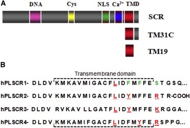
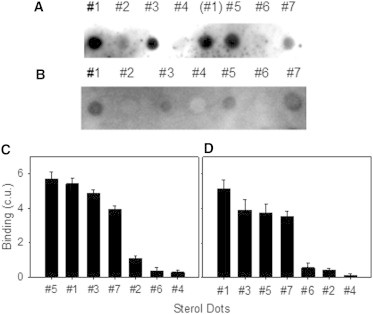
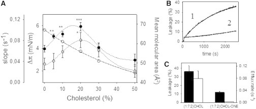
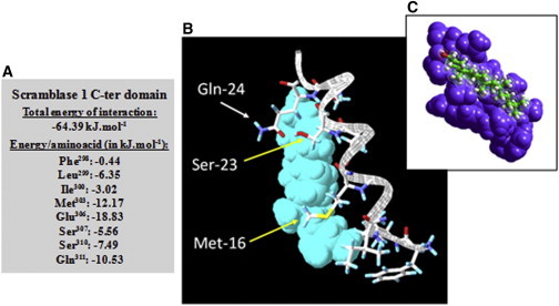
Human phospholipid scramblase 1 (SCR) catalyzes phospholipid transmembrane (flip-flop) motion. This protein is assumed to bind the membrane hydrophobic core through a transmembrane domain (TMD) as well as via covalently bound palmitoyl residues. Here, we explore the possible interaction of the SCR TMD with cholesterol by using a variety of experimental and computational biophysical approaches. Our findings indicate that SCR contains an amino acid segment at the C-terminal region that shows a remarkable affinity for cholesterol, although it lacks the CRAC sequence. Other 3-OH sterols, but not steroids lacking the 3-OH group, also bind this region of the protein. The newly identified cholesterol-binding region is located partly at the C-terminal portion of the TMD and partly in the first amino acid residues in the SCR C-terminal extracellular coil. This finding could be related to the previously described affinity of SCR for cholesterol-rich domains in membranes.


Figures

Figure 1
Figure 1
(A) SCR domains and sequence-derived peptides: DNA-binding domain (pink), Cys-rich domain (yellow), nuclear localization signal (green), calcium-binding domain (blue), and TMD (red). (B) Representation of the human phospholipid (hPL) SCR family putative TMD sequence. hPL SCRs 2, 3, and 4 contain a CRAC motif ([L/V]-[X](1-5)-[Y]-[X](1-5)-[R/K]) localized at the C-terminal end of the predicted transmembrane helix (amino acids in red), whereas SCR presumably is the result of several mutations (in green), such as the polar amino acid (serine) instead of a positively charged amino acid (arginine or lysine), and two phenylalanines in different positions, which appear as tyrosines in the rest of the family sequences. Thus, neither the CRAC nor the CRAC-like sequence ([L/V]-[X](1-5)-[F]-[X](1-5)-[R/K]) is completed in SCR TMD. Sequence information was taken from UNIPROT. Sequences were aligned according to Sahu et al. (3) and Bateman et al. (7).
Figure 2
Figure 2
Solid-phase binding assay. Dot blots of TM31C (A) and SCR (B) binding to Chol (#1) and different analogs: cholestanone (#2), ergosterol (#3), 5α-cholestane (#4), 7-dehydrocholesterol (#5), Chol-one (#6), and coprostanol (#7). (C and D) The results are presented in decreasing order of peptide (C) and SCR (D) binding to sterol analogs. The binding indicates the importance of the alcohol group in ring A for interaction with the scramblase TMD.
Figure 3
Figure 3
(A) Surface pressure measurements of TM31C in PC/Chol monolayers. The increase in surface pressure (●) is shown after insertion of TM31C into lipid monolayers (π0 = 24 ± 0.5 mN/m) made of PC and increasing concentrations of Chol; the calculated slopes from the time traces are represented by ○, dotted line. Experimental data are fitted to a polynomial function (degree = 2). Symbols correspond to the mean ± SDs of four to six independent measurements. The mean molecular areas (○, dashed line) were taken from Li et al. (29) to show the increased monolayer packing. (B and C) ANTS-DPX vesicle content efflux. (B) Time course of TMC31-induced release of intravesicular aqueous contents for POPC/PSM/Chol (1) and POPC/PSM/Chol-one (2) 1:7:2 composition. (C) TM31C-induced total release (black bars) and corresponding initial rates (white bars).
Figure 4
Figure 4
(A and B) DSC of pure SOPC/Chol 1:1 (mol:mol) (thermogram 1) and the same lipid mixtures with 2.5 mol % (2A or 2B), 5 mol % (3A or 3B), and 7.5 mol % (4A or 4B) TM31C or TM19, respectively. Scans are offset along the y axis for clarity of presentation. The scan rate was 1.5°C/min from 10°C to 50°C. The lipid concentration was 4 mM in 20 mM PIPES (pH 7.4), 150 mM NaCl, 1 mM EDTA. (C) Heating-cooling scans of 5 mol % TM31C- (thermograms 1 and 2) or TM19- (thermograms 3 and 4) induced changes when inserted into SOPC/Chol 1:1 (mol ratio). (D) DSC measurements of pure SOPC (thermogram 1) and mixtures with 7.5 mol % TM31C (thermogram 2) or TM19 (thermogram 3). Only cooling scans are shown in D.
Figure 5
Figure 5
Docking of Chol on hPL SCR. A short part of TM31C, [298FLIDFMFFESTGSQ311], contains the highest enthalpic interaction with Chol. (A) Detailed analysis of the energetics of Chol interaction with the protein TMD and other surrounding amino acids that presumably are involved in the interaction. (B) Surface view of the complex, with important amino acid side chains presented as interacting with the sterol. The TMD amino acid M303 is represented as Met-16, which interacts with the Chol ring structure, and the C-terminal coil amino acids S309 and Q311, which interact with the small polar alcohol headgroup, are represented as Ser-23 and Gln-24 respectively. E306, at the end of TMD (which is not represented), contains the highest enthalpic interaction with Chol. (C) Overall illustration of the interaction of scramblase TMD C-ter coil (in blue) and Chol.
Figure 6
Figure 6
(A) Dot blots of the fractions recovered from the sucrose gradient (27) (fractions 1–4, samples recovered from top to bottom of the sucrose gradient). The yield of protein reconstitution into PC/SM/Chol 2:1:1 and 1:1:1 vesicles was virtually the same (∼50% of SCR successfully reconstituted). (B) SCR quantification in each recovered fraction when reconstituted in PC/SM/Chol 2:1:1 vesicles (gray bars) or PC/PS 9:1 vesicles (black bars). The recovered top fraction was used for the flip-flop assay. White bars correspond to pure ultracentrifuged protein. (C and D) Influence of Chol on py-SM (C) and NBD-PS (D) transbilayer movement promoted by SCR. All measurements were made in the presence of 5 mM calcium. (Δ) Liposomes. (▲) SCR proteoliposomes of DOPC/SM/Chol 2:1:1. (▪) SCR proteoliposomes of POPC/PS/Chol 9:1:3.3. (●) SCR proteoliposomes of PC/PS 9:1. (○) SCR proteoliposomes in the absence of Ca2+. Average values ± SEM (n ≥ 3).
Figure 7
Figure 7
Suggested outline of the SCR C-terminal domain disposition in membranes when inserted into Chol-depleted domains (left) and Chol-rich domains (right). Red, extracellular segment; green, the putative TMD; blue, the cytoplasmic portion.

References

    1. Bassé F., Stout J.G., Wiedmer T. Isolation of an erythrocyte membrane protein that mediates Ca2+-dependent transbilayer movement of phospholipid. J. Biol. Chem. 1996;271:17205–17210. - PubMed
    1. Zhou Q., Zhao J., Sims P.J. Molecular cloning of human plasma membrane phospholipid scramblase. A protein mediating transbilayer movement of plasma membrane phospholipids. J. Biol. Chem. 1997;272:18240–18244. - PubMed
    1. Sahu S.K., Gummadi S.N., Aradhyam G.K. Phospholipid scramblases: an overview. Arch. Biochem. Biophys. 2007;462:103–114. - PubMed
    1. Ben-Efraim I., Zhou Q., Sims P.J. Phospholipid scramblase 1 is imported into the nucleus by a receptor-mediated pathway and interacts with DNA. Biochemistry. 2004;43:3518–3526. - PubMed
    1. Wyles J.P., Wu Z., Cole S.P. Nuclear interactions of topoisomerase II alpha and beta with phospholipid scramblase 1. Nucleic Acids Res. 2007;35:4076–4085. - PMC - PubMed

Publication types