. 2014 Sep 16;107(6):1403-14.
doi: 10.1016/j.bpj.2014.07.055.

In vivo orientation of single myosin lever arms in zebrafish skeletal muscle

Affiliations

In vivo orientation of single myosin lever arms in zebrafish skeletal muscle

Xiaojing Sun et al. Biophys J. .

Abstract

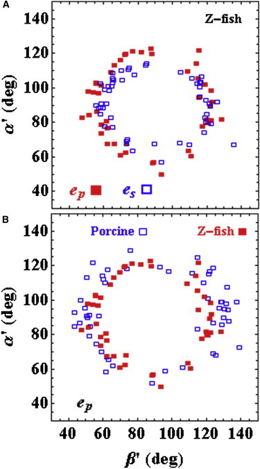
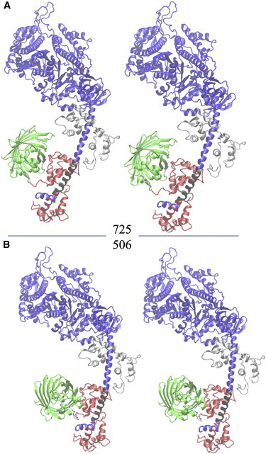
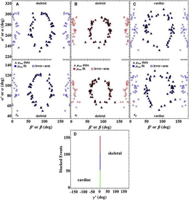
Cardiac and skeletal myosin assembled in the muscle lattice power contraction by transducing ATP free energy into the mechanical work of moving actin. Myosin catalytic/lever-arm domains comprise the transduction/mechanical coupling machinery that move actin by lever-arm rotation. In vivo, myosin is crowded and constrained by the fiber lattice as side chains are mutated and otherwise modified under normal, diseased, or aging conditions that collectively define the native myosin environment. Single-myosin detection uniquely defines bottom-up characterization of myosin functionality. The marriage of in vivo and single-myosin detection to study zebrafish embryo models of human muscle disease is a multiscaled technology that allows one-to-one registration of a selected myosin molecular alteration with muscle filament-sarcomere-cell-fiber-tissue-organ- and organism level phenotypes. In vivo single-myosin lever-arm orientation was observed at superresolution using a photoactivatable-green-fluorescent-protein (PAGFP)-tagged myosin light chain expressed in zebrafish skeletal muscle. By simultaneous observation of multiphoton excitation fluorescence emission and second harmonic generation from myosin, we demonstrated tag specificity for the lever arm. Single-molecule detection used highly inclined parallel beam illumination and was verified by quantized photoactivation and photobleaching. Single-molecule emission patterns from relaxed muscle in vivo provided extensive superresolved dipole orientation constraints that were modeled using docking scenarios generated for the myosin (S1) and GFP crystal structures. The dipole orientation data provided sufficient constraints to estimate S1/GFP coordination. The S1/GFP coordination in vivo is rigid and the lever-arm orientation distribution is well-ordered in relaxed muscle. For comparison, single myosins in relaxed permeabilized porcine papillary muscle fibers indicated slightly differently oriented lever arms and rigid S1/GFP coordination. Lever arms in both muscles indicated one preferred spherical polar orientation and widely distributed azimuthal orientations relative to the fiber symmetry axis. Cardiac myosin is more radially displaced from the fiber axis. Probe rigidity implies the PAGFP tag reliably indicates cross-bridge orientation in situ and in vivo.


Figures

Figure 1
Figure 1
The PDMS microfluidic on a glass coverslip forming the zebrafish embryo confinement chamber. (A) Schematic side view of the chamber showing its aqueous, PDMS, and glass components and depth. (B) The actual device and confined embryo in top view as it would be imaged by the 2-P upright microscope objective. Inlet and outlet ports are for solution exchange. (C) A PDMS cube creates a cavity filled with an aqueous solution containing fluorescein, imitating the fish medium in a microfluidic channel. The HILO beam is created by an oil immersion objective (100×, 1.3 NA, and 200 μm working distance) and emerges into the aqueous side of the interface at ∼74°.
Figure 2
Figure 2
The excitation pathway schematic for HILO illumination. Incident (i), reflected (r), and transmitted (t) beams in blue have s- or p-polarization (eˆsoreˆp) for the transmitted beam. They are parallel or perpendicular to the apparent symmetry axis of the in vivo skeletal muscle fiber. Lab fixed coordinates are of two types, TIRF coordinates (θ,ϕ) relative to (Lx,Ly,Lz) and fiber coordinates (β′,α′) relative to (Lx,Ly,Lz), depicting the orientation of the (photoactivated) emission dipole, μˆe. Symbol σ is the incidence angle of the exciting beam. To see this figure in color, go online.
Figure 3
Figure 3
Simultaneous RLC-GFP fluorescence and myosin SHG in vivo by 2-P excitation. Infrared light at 810 nm excites fluorescence at ∼500 nm (green) and SHG at 405 nm (blue) in the wide view of the trunk skeletal muscles in the tail (left) and in the close-up of two individual fibers (right). The I-band, with actin but no myosin, and the M-line, without myosin cross bridges, are dark in comparison to the A-band, which contains both myosin cross bridges and actin. The A-band shows both GFP fluorescence and SHG signals indicating colocalization of RLC-GFP with the cross bridge. Sarcomere length is ∼2.1 μm.
Figure 4
Figure 4
In vivo images of RLC-PAGFP- (upper) to RLC-GFP-tagged myosin (lower) from zebrafish embryos in relaxation under HILO illumination.
Figure 5
Figure 5
Zebrafish skeletal and porcine cardiac muscle data from relaxed fibers. (A) Comparison of zebrafish skeletal myosin data for p- and s-polarized photoactivation. (B) Comparison of data for p-polarized photoactivation porcine cardiac myosin and zebrafish skeletal myosin.
Figure 6
Figure 6
Stereo representations of S1/GFP. The myosin sequence shows the MHC (blue or black), the RLC (red), the GFP (green), and the ELC (silver). The 11-residue sequence linking the RLC C terminus to the GFP N terminus is white. The black section of the MHC where RLC binds to the lever arm defines an α-helix symmetry axis with spherical polar coordinates (β,α) defined relative to the lab coordinates (Lx,Ly,Lz) in Fig. 2. (A) Structure 725 is the best S1/GFP representation of fish skeletal myosin. (B) Structure 506 is the best S1/GFP representation of porcine cardiac myosin.
Figure 7
Figure 7
Scatter plots for single emission-dipole and lever-arm orientation coordinates, (β′,α′) or (β,α), respectively, from skeletal (A and B) and cardiac muscle (C) in relaxation. Blue and red symbols represent p-polarized (ep) and s-polarized (es) photoactivating light polarization. Open squares (lever-arm) are calculated from the emission dipole moments of photoactivated probes with orientation coordinates (β′,α′) (open triangles). The fitted points (solid blue and red squares (μemfit)) were derived from structures 725 (skeletal) and 506 (cardiac) in Fig. 6. (D) Stacked histogram for the absorption dipole coordinate, γ′, defined in Eq. 12d. The histogram specifies one of the Euler angles relating the emission molecular frame ({u,v,w} in Fig. S1) to the lab frame ({Lx,Ly,Lz} in Fig. 2). The narrow-profile histogram indicates that PAGFP is rigidly immobilized on the cross bridge in both skeletal and cardiac muscle. The skeletal histogram has events from both p- and s-polarization (blue and red, respectively) photoactivated dipoles. The cardiac histogram has just the p-polarization photoactivated dipoles (green).

References

    1. Lowey S., Waller G.S., Trybus K.M. Function of skeletal muscle myosin heavy and light chain isoforms by an in vitro motility assay. J. Biol. Chem. 1993;268:20414–20418. - PubMed
    1. Sherwood J.J., Waller G.S., Lowey S. A point mutation in the regulatory light chain reduces the step size of skeletal muscle myosin. Proc. Natl. Acad. Sci. USA. 2004;101:10973–10978. - PMC - PubMed
    1. Pant K., Watt J., Moore J.R. Removal of the cardiac myosin regulatory light chain increases isometric force production. FASEB J. 2009;23:3571–3580. - PMC - PubMed
    1. Oldfors A. Hereditary myosin myopathies. Neuromuscul. Disord. 2007;17:355–367. - PubMed
    1. Toydemir R.M., Rutherford A., Bamshad M.J. Mutations in embryonic myosin heavy chain (MYH3) cause Freeman-Sheldon syndrome and Sheldon-Hall syndrome. Nat. Genet. 2006;38:561–565. - PubMed

Publication types

LinkOut - more resources