. 2014 Sep 16;107(6):L09-12.
doi: 10.1016/j.bpj.2014.07.054.

Real-time imaging of electrical signals with an infrared FDA-approved dye

Affiliations

Real-time imaging of electrical signals with an infrared FDA-approved dye

Jeremy S Treger et al. Biophys J. .

Abstract

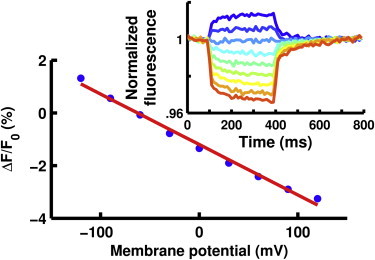
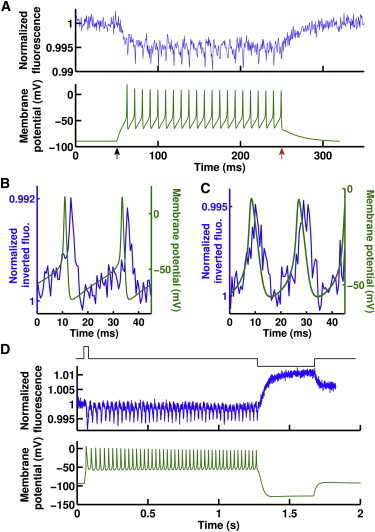
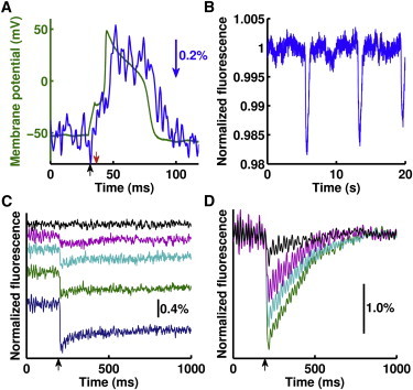
Clinical methods used to assess the electrical activity of excitable cells are often limited by their poor spatial resolution or their invasiveness. One promising solution to this problem is to optically measure membrane potential using a voltage-sensitive dye, but thus far, none of these dyes have been available for human use. Here we report that indocyanine green (ICG), an infrared fluorescent dye with FDA approval as an intravenously administered contrast agent, is voltage-sensitive. The fluorescence of ICG can follow action potentials in artificial neurons and cultured rat neurons and cardiomyocytes. ICG also visualized electrical activity induced in living explants of rat brain. In humans, ICG labels excitable cells and is routinely visualized transdermally with high spatial resolution. As an infrared voltage-sensitive dye with a low toxicity profile that can be readily imaged in deep tissues, ICG may have significant utility for clinical and basic research applications previously intractable for potentiometric dyes.


Figures

Figure 1
Figure 1
ICG-labeled oocytes showed that ICG’s fluorescence (blue points) is roughly linearly dependent (red line, fit to data) with voltage. (Inset) Oocyte membrane potential was held at −60 mV and then pulsed to potentials ranging from −120 mV (blue) to +120 mV (red). Ex: 780 nm, Em: 818–873 nm.
Figure 2
Figure 2
ICG can monitor action potentials. (A) Oocytes coinjected with voltage-gated sodium and potassium channel cRNA fired action potentials (bottom, green) when held under current clamp. ICG fluorescence changes (top, blue) detected these action potentials at a rate of 107 Hz. Stimulus start (black arrow) and end (red arrow) are shown. (B and C) ICG fluorescence (blue, inverted) distinguished between healthy action potentials from wild-type sodium channels (B, green) and diseased action potentials from sodium channels with a myotonic substitution (C, green). Cells are stimulated for the entire time course of these panels. The delay between action potentials and the ICG signal is due to a low-pass filtering effect caused by the dye response time and the camera integration time. (D) In cells with myotonic sodium channels, a brief stimulus (top, black) was sufficient to elicit a train of action potentials (bottom, green) that only ceased upon significant hyperpolarization, as expected in a myotonia. ICG fluorescence (middle, blue) successfully followed each one of these action potentials.
Figure 3
Figure 3
ICG follows electrical activity in living mammalian tissue. (A) Rat cultured dorsal root ganglion cells under current-clamp (black arrow, pulse start; red arrow, pulse end) fired action potentials (green), that ICG fluorescence tracked (blue, inverted, low-pass-filtered at 225 Hz; blue arrow, relative fluorescence change). (B) ICG fluorescence sensed spontaneous membrane potential changes in cardiomyocyte syncytia. (C) In rat brain slices, ICG responds differently to no stimulus (black) and stimuli of increasing intensity (magenta, cyan, green, and blue, increasing amplitude; scale bar shows relative fluorescence change). Weaker stimuli traces (e.g., magenta) show complete fluorescence recovery whereas larger stimuli (e.g., blue) do not fully recover within this time course; traces are vertically offset for clarity. (D) Tetrodotoxin (TTX) reduced the ICG response to a stimulus over 12 min (green, pre-TTX; cyan, magenta, and black, increasing time post-TTX; low-pass-filtered at 40 Hz; black arrow, stimulus).

References

    1. Cohen L.B., Salzberg B.M. Optical measurement of membrane potential. Rev. Physiol. Biochem. Pharmacol. 1978;83:35–88. - PubMed
    1. Peterka D.S., Takahashi H., Yuste R. Imaging voltage in neurons. Neuron. 2011;69:9–21. - PMC - PubMed
    1. Shapiro M.G., Priest M.F., Bezanilla F. Thermal mechanisms of millimeter wave stimulation of excitable cells. Biophys. J. 2013;104:2622–2628. - PMC - PubMed
    1. Buzsáki G., Mizuseki K. The log-dynamic brain: how skewed distributions affect network operations. Nat. Rev. Neurosci. 2014;15:264–278. - PMC - PubMed
    1. Lerche H., Heine R., Lehmann-Horn F. Human sodium channel myotonia: slowed channel inactivation due to substitutions for a glycine within the III-IV linker. J. Physiol. 1993;470:13–22. - PMC - PubMed

Publication types