. 2014 Oct 30;5(20):9783-97.
doi: 10.18632/oncotarget.2392.

Systematic dissection of the mechanisms underlying progesterone receptor downregulation in endometrial cancer

Affiliations

Systematic dissection of the mechanisms underlying progesterone receptor downregulation in endometrial cancer

Shujie Yang et al. Oncotarget. .

Abstract

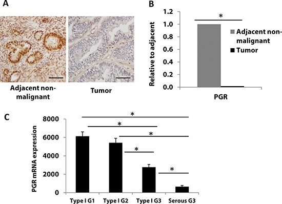
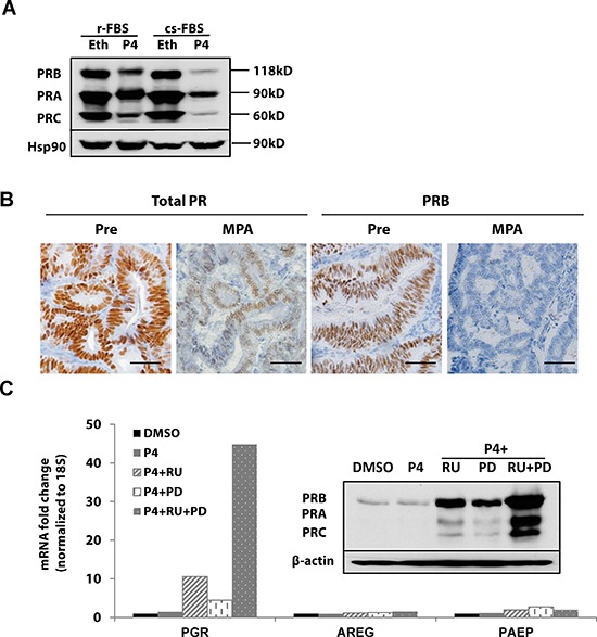
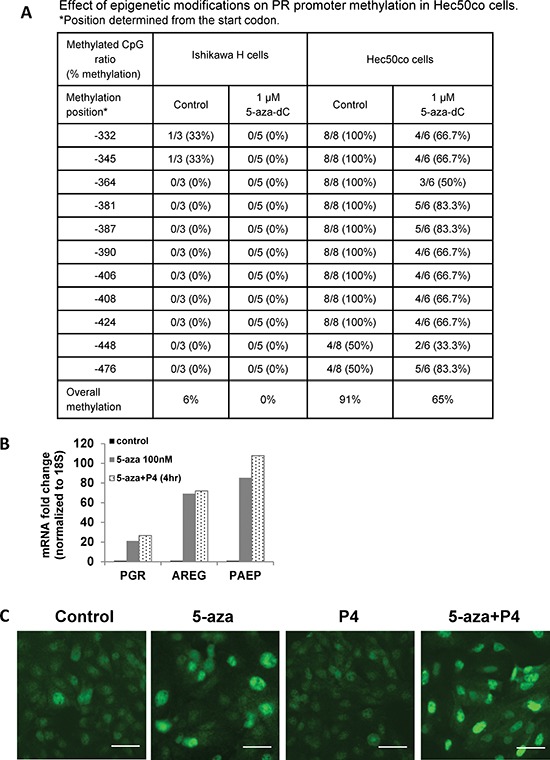
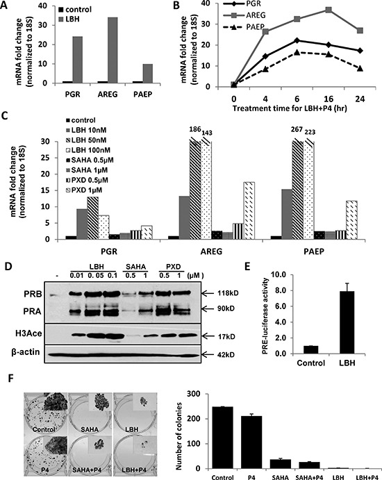
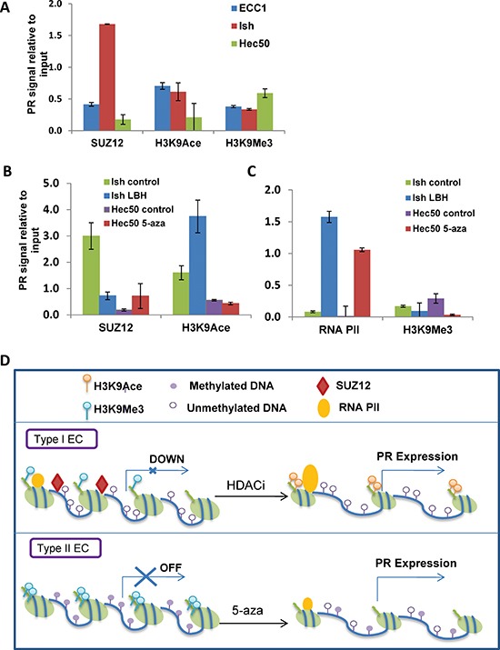
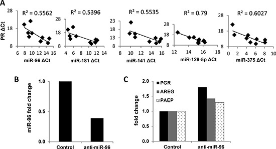
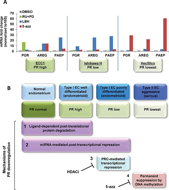
Progesterone, acting through its receptor, PR (progesterone receptor), is the natural inhibitor of uterine endometrial carcinogenesis by inducing differentiation. PR is downregulated in more advanced cases of endometrial cancer, thereby limiting the effectiveness of hormonal therapy. Our objective was to understand and reverse the mechanisms underlying loss of PR expression in order to improve therapeutic outcomes. Using endometrial cancer cell lines and data from The Cancer Genome Atlas, our findings demonstrate that PR expression is downregulated at four distinct levels. In well-differentiated cancers, ligand-induced receptor activation and downregulation are intact. miRNAs mediate fine tuning of PR levels. As differentiation is lost, PR silencing is primarily at the epigenetic level. Initially, recruitment of the polycomb repressor complex 2 to the PR promoter suppresses transcription. Subsequently, DNA methylation prevents PR expression. Appropriate epigenetic modulators reverse these mechanisms. These data provide a rationale for combining epigenetic modulators with progestins as a therapeutic strategy for endometrial cancer.

Significance: Traditional hormonal therapy for women with endometrial cancer can be molecularly enhanced by combining progestins with epigenetic modulators, thereby increasing progesterone receptor expression and significantly improving treatment efficacy.


Figures

Figure 1
Figure 1. Progesterone receptor expression is frequently downregulated with progression of endometrial cancer
A, B PR protein (A), and PGR mRNA (B), expression was measured in endometrial tumors (n=5) and matched adjacent non-malignant tissue (n=5) by immunostaining and real-time PCR, respectively. Scale bar = 50 μm. Student's t-test was used for comparisons of two groups. (C) PGR mRNA expression was analyzed in 361 endometrial cancer patient tumors from TCGA database. Patients were divided into four groups: endometrioid type I grade 1 (G1, n=84), grade 2 (G2, n=100), grade 3 (G3, n=115) and serous Type II grade 3 (G3, n=62). Error bars, SEM. Statistical analysis was conducted using one way ANOVA with significance level set at α=0.05. All pairwise multiple comparisons were performed using Holm-Sidak method with Bonferroni correction. The results showed that all individual groups are significantly different from each other (p< 0.001) except between Type I G1 and Type I G2 (p=0.209).
Figure 2
Figure 2. Ligand-dependent PR downregulation
(A) Western blotting: T47D breast cancer cells were grown in DMEM media supplemented with regular fetal bovine serum (r-FBS) or charcoal-stripped serum (cs-FBS), followed by treatment with ethanol (vehicle control) or 100 nM progesterone (P4) for 24h. PR protein was detected using specific PR antibodies, and HSP90 serves as a loading control. (B) Immunohistochemistry: endometrial tumor specimens were collected before or 21 days after MPA treatment (400 mg, intramuscularly) and total PR and PRB expression was assessed by immunohistochemistry. Scale bar = 50 μm. (C) q-PCR and Western blotting: ECC1 cells were treated with DMSO (vehicle control), 100 nM P4, 100 nM P4 and 1μM PR antagonist RU486 (RU), 100 nM P4+ 1 μM MAPK inhibitor PD0325901(PD) or the combination of P4+RU+PD for 24h. mRNA expression of PGR, AREG and PAEP was measured by q-PCR, normalized to 18S, and data displayed as fold-change relative to DMSO control. Comparisons of normalized expression values (ΔCt) employed the conventional ΔΔCt fold change method. The insert is PR protein expression after the same treatment; β-actin, loading control.
Figure 3
Figure 3. PGR promoter methylation represses PR expression in serous endometrial cancer cells
(A) Bisulfite sequencing. PGR promoter methylation was quantified in Ishikawa H and Hec50co cells before or after hypomethylating agent 5-aza-decitabine (5-aza-dC) treatment. (B) q-PCR analysis. Hec50co cells were treated with DMSO (vehicle control), 100 nM 5-aza-dC for 5 days with fresh 5-aza-dC added every other day, 5-aza-dC for 5 days +100 nM P4 for 4h. mRNA expression of PGR, AREG and PAEP was measured by q-PCR and normalized to 18S, and data are displayed as fold-change relative to DMSO control. Comparisons of normalized expression values (ΔCt) employed the conventional ΔΔCt fold change method. (C) Representative immunofluorescent image showing PR expression in Hec50co cells treated with DMSO control, 5 days of 100 nM 5-aza-dC, 100 nM P4, or both. Scale bar = 50 μm.
Figure 4
Figure 4. Histone deacetylase (HDAC) inhibition restores PR mRNA and protein expression in Type I Ishikawa H cells
(A) q-PCR analysis. Ishikawa H cells were treated with DMSO control or 20 nM LBH589 (LBH) for 24 h. PGR, AREG and PAEP mRNA expression was normalized to 18S, and all q-PCR data are displayed as fold-change relative to DMSO control. Comparisons of normalized expression values (ΔCt) employed the conventional ΔΔCt fold change method. (B) PR expression and activity corresponding to time course of P4 stimulation: Ishikawa H cells were treated with 20 nM LBH +100 nM P4 for the indicated times, and PGR, AREG and PAEP mRNA expression was quantified by q-PCR and normalized to 18S. (C) q-PCR analysis. Ishikawa H cells were treated with three different HDAC inhibitors (LBH589, SAHA and PXD101) at the indicated concentrations for 24h. PGR, AREG and PAEP mRNA expression was quantified by q-PCR and normalized to 18S. (D) Western blotting. Expression of PR protein in Ishikawa H was measured after treating with the three HDAC inhibitors. The presence of histone H3 acetylation indicates drug effect, and β-actin serves as a loading control. (E) PRE-luciferase assay. Ishikawa H cells were treated with 20 nM LBH589 for 24h and studied using a PRE-luciferase assay. The PRE-luciferase activity was normalized to total protein concentration. (F) Colony formation assay. Ishikawa H cells were treated in the presence or absence of HDACi for 2 weeks, and resulting colonies were stained with crystal violet (left panel, insets are 5X) and the number of colonies recorded (right panel). Error bar, SD.
Figure 5
Figure 5. Mechanisms of progressive PR silencing in endometrial cancer cells
(A) ChIP followed by q-PCR analysis of SUZ12, H3K9Ace or H3K9Me3 recruitment to the PGR promoter. (B) ChIP-PCR analysis. Ishikawa H cells were treated with or without 20 nM LBH589 for 24h, and Hec50 cells were treated with or without the hypomethylating agent 100 nM 5-aza-deoxycytidine (5-aza) for 5 days. ChIP followed by q-PCR for SUZ12 and H3K9Ace was used to determine recruitment of these factors to the PGR promoter. (C) ChIP-PCR analysis. ChIP followed by q-PCR for RNA polymerase II (RNA PII) and H3K9 trimethylation (H3K9Me3) was performed to assess occupancy on the PGR promoter. (D) Proposed model for PR repression in well-differentiated and poorly-differentiated endometrial cancer. In well-differentiated endometrial cancers, PR was transcriptional repressed by PRC2 and reversed by HDACi treatment, while in poorly-differentiated endometrial cancers, PR was suppressed by DNA methylation and reversed by a hypomethylating agent.
Figure 6
Figure 6. Impact of miRNAs on PR expression in endometrial tumors and cells
(A) Correlation of miRNA expression with PGR expression in matched non-malignant endometrial tissue vs. endometrioid adenocarcinoma (n=5). mRNA expression of PGR was measured by q-PCR and normalized to 18S and miRNA expression was measured by q-PCR and normalized to RNU48, and both data are displayed as ΔCt value relative to the DMSO control. (B) miR-96 was decreased by transfecting the miR-96 inhibitor into Ishikawa H cells. (C) PR expression was restored by inhibiting miR-96 in Ishikawa H cells. Comparisons of normalized miRNA expression values (ΔCt) employed the conventional ΔΔCt fold change method.
Figure 7
Figure 7. Systematic analysis of strategies to restore functional PR expression in distinct models of endometrial cancer
(A) Three endometrial cancer model cell lines, ECC1, Ishikawa H and Hec50co cells were treated with DMSO control, 1 μM RU486+1 μM PD0325901 for 24h, 20 nM LBH589 for 24h or 100 nM 5-aza for 3 days. mRNA expression of PGR, AREG and PAEP were measured by q-PCR, normalized to 18S and displayed as fold change to DMSO control. Comparisons of normalized expression values (ΔCt) employed the conventional ΔΔCt fold change method. In ECC1 cells, which express PR at baseline, treatment with the progestin antagonist RU486 and the MAPK inhibitor PD0325901 validates the dynamic PR regulation by ligand-mediated degradation. In Type I Ishikawa cells, only an HDACi effectively restores functional PR, whereas a hypomethylating agent is necessary for Type II Hec50co cells. (B) Proposed model of PR downregulation mechanisms during endometrial cancer progression.

References

    1. Siegel R, Naishadham D, Jemal A. Cancer statistics, 2013. CA Cancer J Clin. 2013;63(1):11–30. - PubMed
    1. Yang S, Thiel KW, Leslie KK. Progesterone: the ultimate endometrial tumor suppressor. Trends Endocrinol Metab. 2011;22(4):145–152. - PMC - PubMed
    1. Kim JJ, Chapman-Davis E. Role of progesterone in endometrial cancer. Seminars in Reproductive Medicine. 2010;28(01):081–090. - PMC - PubMed
    1. Singh M, Zaino RJ, Filiaci VJ, Leslie KK. Relationship of estrogen and progesterone receptors to clinical outcome in metastatic endometrial carcinoma: a Gynecologic Oncology Group Study. Gynecol Oncol. 2007;106(2):325–333. - PubMed
    1. Dahmoun M, Backstrom T, Boman K, Cajander S. Apoptosis, proliferation, and hormone receptors in endometrial carcinoma: results depending on methods of analysis. Int J Oncol. 2003;22(1):115–122. - PubMed

Publication types

MeSH terms