Review
. 2015 Jan;235(2):242-52.
doi: 10.1002/path.4445.

Molecular biology, pathogenesis and pathology of mumps virus

Affiliations
Review

Molecular biology, pathogenesis and pathology of mumps virus

Steven Rubin et al. J Pathol. 2015 Jan.

Abstract

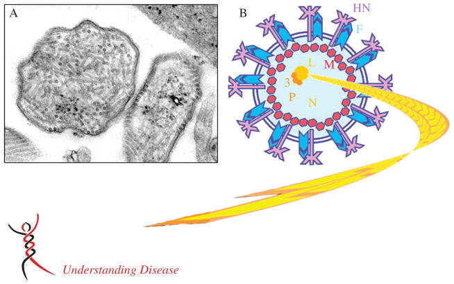
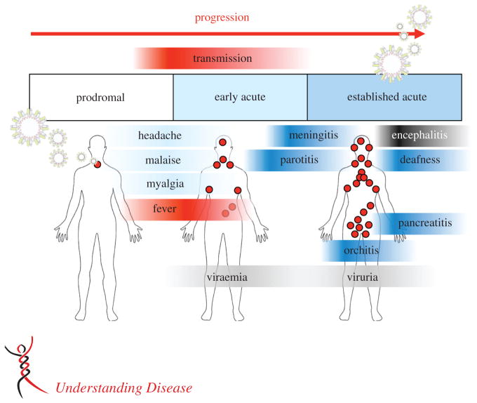
Mumps is caused by the mumps virus (MuV), a member of the Paramyxoviridae family of enveloped, non-segmented, negative-sense RNA viruses. Mumps is characterized by painful inflammatory symptoms, such as parotitis and orchitis. The virus is highly neurotropic, with laboratory evidence of central nervous system (CNS) infection in approximately half of cases. Symptomatic CNS infection occurs less frequently; nonetheless, prior to the introduction of routine vaccination, MuV was a leading cause of aseptic meningitis and viral encephalitis in many developed countries. Despite being one of the oldest recognized diseases, with a worldwide distribution, surprisingly little attention has been given to its study. Cases of aseptic meningitis associated with some vaccine strains and a global resurgence of cases, including in highly vaccinated populations, has renewed interest in the virus, particularly in its pathogenesis and the need for development of clinically relevant models of disease. In this review we summarize the current state of knowledge on the virus, its pathogenesis and its clinical and pathological outcomes.

Keywords: mumps; mumps virus; neurovirulence; pathogenesis; vaccine.


Conflict of interest statement

No conflicts of interest were declared.

Figures

Figure 1
Figure 1
MuV virion structure. (A) Thin sectioned transmission electron micrograph showing a typical MuV particle alongside (B) a schematic of the particle. The enveloped particles are pleomorphic, in the size range 100–600 nm. Within this structure lies the long, coiled electron-dense ribonucleoprotein (RNP), containing the MuV genome. Small spikes can be observed on the surface of the particle, corresponding to the viral HN and F glycoproteins. The same general features of the MuV particle are shown in the schematic (B). The envelope (blue lines) is studded with the HN (purple) and F (blue) glycoproteins and encases the viral RNP, made up of the RNA genome (3′–5′) in complex with N (yellow), P (orange) and L (gold) proteins. The M protein (red) interacts with the envelope, glycoproteins and the RNP. The V, I and SH proteins are expressed in infected cells, but are not thought to be incorporated within the virion. Photomicrograph courtesy of CDC/A Harrison and FA Murphy (http://phil.cdc.gov/phil/details.asp)
Figure 2
Figure 2
MuV clinical presentation and pathogenesis. Mumps is a respiratory-spread, acute, inflammatory disease in humans, which causes a range of systemic symptoms. The incubation period is 2–4 weeks. Approximately one-third of infections are asymptomatic. The prodromal phase is characterized by non-specific, often mild symptoms, such as low-grade fever, headache and malaise. An early acute phase follows, likely representing spread of the virus from the respiratory tract and development of systemic symptoms, typically parotitis, which lasts from a few days to 1 week. During the established acute phase, orchitis, meningitis or encephalitis may appear. Symptoms usually resolve within 2 weeks, coincident with the development of a MuV-specific humoral response. Long-term complications and death are rare
Figure 3
Figure 3
MuV infection of the rat brain. The most prominent neuropathological outcome following MuV intracranial inoculation in small animal models (hamsters, rats) is enlargement of the lateral and third ventricles, ie hydrocephalus, which has also been reported in cases in humans. The cause of hydrocephalus is postulated to be denuding of virus-infected ependymal cells lining the ventricles. (A) Sagittal section of rat brain tissue immunohistochemically stained for the MuV nucleoprotein, showing extensive infection of the ventricular ependymal cells (green foci). (B) Approximately 3 weeks later, ependymal cell loss is evident in comparison to the well-preserved ependymal cell architecture in rats injected with the Jeryl Lynn vaccine strain (C); haematoxylin and eosin (H&E) stain
Figure 4
Figure 4
Hydrocephalus severity in MuV-infected rats. (A) H&E-stained sagittal sections of brain from a representative 30 day-old rat injected with a wild-type (WT) MuV isolate as a newborn (top), compared to a rat similarly injected with the highly attenuated Jeryl Lynn (JL) vaccine strain (bottom). (B) T1 weighted gradient-echo image from MRI of the same brains as in (A) (upper left corner and lower right corner), compared to brain from a rat injected with an insufficiently attenuated vaccine strain, Urabe-AM9 (Ur, lower left corner) and an uninfected rat brain (0, upper right corner). Note that the severity of hydrocephalus tracks with the virus strain’s neurovirulence potential for humans. (C) Assembled three-dimensional rendering of MRI slices represented in (B), showing ventricular volume (blue, wild-type; pink, Urabe-AM9)
Figure 5
Figure 5
A number of important questions remain unresolved regarding MuV pathogenesis. This is of particular relevance to renewed efforts towards development of a more efficacious MuV vaccine, in light of the resurgence of mumps in vaccinated populations. The classic method of virus attenuation is extensive blind passage in vitro. While this often leads to the desired effect of a loss of virulence and reactogenicity, it can also lead to loss of immunogenicity and efficacy. Clearly, a more rational approach to virus attenuation is needed, and understanding the natural pathogenesis of the infectious agent is a prerequisite to any such endeavour. This figure highlights this issue, showing our current assumptions of pathogenesis (black text) and unresolved questions (red text)

References

    1. Wagenvoort JH, Harmsen M, Boutahar-Trouw BJ, et al. Epidemiology of mumps in The Netherlands. J Hyg (Lond) 1980;85:313–326. - PMC - PubMed
    1. Anderson RM, Crombie JA, Grenfell BT. The epidemiology of mumps in the UK: a preliminary study of virus transmission, herd immunity and the potential impact of immunization. Epidemiol Infect. 1987;99:65–84. - PMC - PubMed
    1. Cox MJ, Anderson RM, Bundy DA, et al. Seroepidemiological study of the transmission of the mumps virus in St. Lucia, West Indies. Epidemiol Infect. 1989;102:147–160. - PMC - PubMed
    1. Centers for Disease Control and Prevention. Current trends mumps – United States, 1984–1985. Morb Mortal Wkly Rep. 1986;35:216–219. - PubMed
    1. McNabb SJ, Jajosky RA, Hall-Baker PA, et al. Summary of notifiable diseases – United States, 2005. Morb Mortal Wkly Rep. 2007;54:1–92. - PubMed

Publication types