. 2015 Feb;113(2):338-49.
doi: 10.1160/TH14-04-0360. Epub 2014 Sep 18.

Antithrombin up-regulates AMP-activated protein kinase signalling during myocardial ischaemia/reperfusion injury

Affiliations

Antithrombin up-regulates AMP-activated protein kinase signalling during myocardial ischaemia/reperfusion injury

Yina Ma et al. Thromb Haemost. 2015 Feb.

Abstract

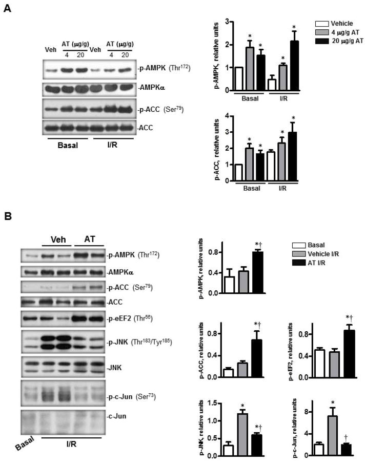
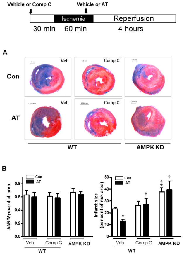
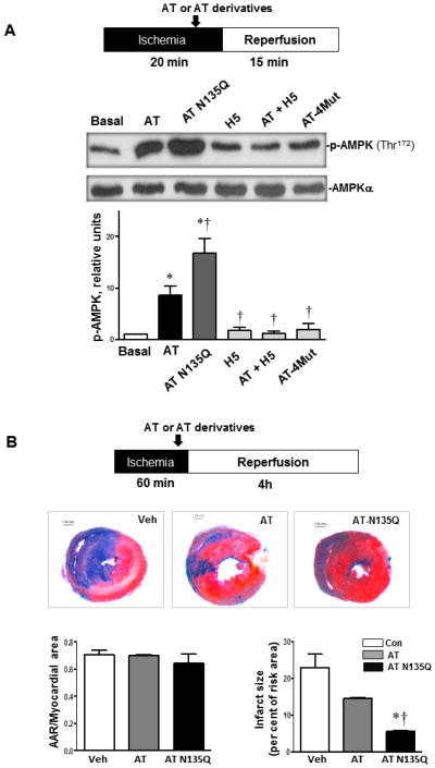
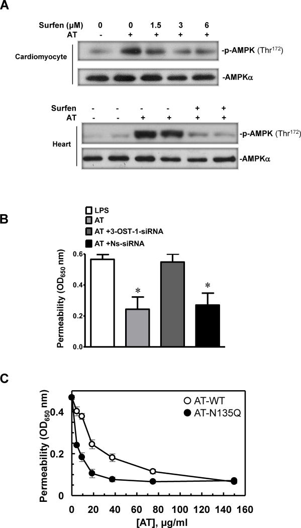
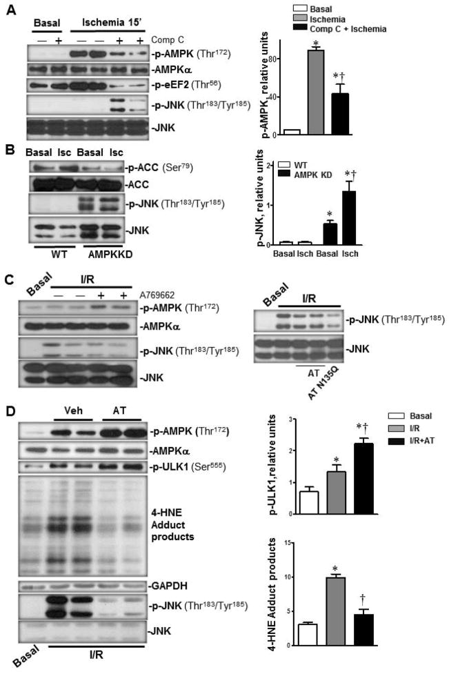
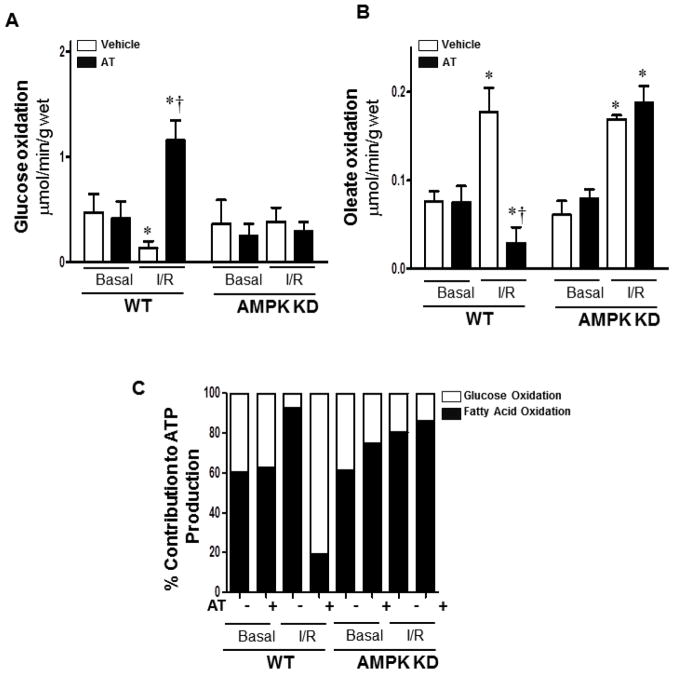
Antithrombin (AT) is a protein of the serpin superfamily involved in regulation of the proteolytic activity of the serine proteases of the coagulation system. AT is known to exhibit anti-inflammatory and cardioprotective properties when it binds to heparan sulfate proteoglycans (HSPGs) on vascular cells. AMP-activated protein kinase (AMPK) plays an important cardioprotective role during myocardial ischaemia and reperfusion (I/R). To determine whether the cardioprotective signaling function of AT is mediated through the AMPK pathway, we evaluated the cardioprotective activities of wild-type AT and its two derivatives, one having high affinity and the other no affinity for heparin, in an acute I/R injury model in C57BL/6J mice in which the left anterior descending coronary artery was occluded. The serpin derivatives were given 5 minutes before reperfusion. The results showed that AT-WT can activate AMPK in both in vivo and ex vivo conditions. Blocking AMPK activity abolished the cardioprotective function of AT against I/R injury. The AT derivative having high affinity for heparin was more effective in activating AMPK and in limiting infraction, but the derivative lacking affinity for heparin was inactive in eliciting AMPK-dependent cardioprotective activity. Activation of AMPK by AT inhibited the inflammatory c-Jun N-terminal protein kinase (JNK) pathway during I/R. Further studies revealed that the AMPK activity induced by AT also modulates cardiac substrate metabolism by increasing glucose oxidation but inhibiting fatty acid oxidation during I/R. These results suggest that AT binds to HSPGs on heart tissues to invoke a cardioprotective function by triggering cardiac AMPK activation, thereby attenuating JNK inflammatory signalling pathways and modulating substrate metabolism during I/R.


Conflict of interest statement

Disclosure of conflict of interests

The authors declare no competing financial interest.

Figures

Figure 1
Figure 1
AT stimulated AMPK activation during in vivo basal and I/R and ex vivo I/R conditions. (A) AT, treated for 20 min at basal condition or 5 min before reperfusion during regional ischemia, induced phosphorylation of AMPK and phosphorylation of downstream acetyl CoA (ACC). The bar graphs (right panel) show the relative level of p-AMPK and p-ACC, respectively. (B) AT-treated (2.5 μg/mL) heart ex vivo at the beginning of reperfusion during global ischemia (25 min). AT increased the phosphorylation of AMPK, ACC and eEF2. AT treatment also inhibited the phosphorylation of JNK and its downstream protein c-Jun during ischemia (25 min) and reperfusion (30 min) (left panel). The bar graphs (right panel) show the relative levels of p-AMPK, p-ACC, p-eEF2, p-JNK and p-c-Jun, respectively. N=5 for basal and I/R condition; n=7 for I/R/AT condition; *p<0.05 vs. Basal Vehicle or I/R Vehicle; †p<0.05 vs. I/R Vehicle.
Figure 2
Figure 2
Activation of AMPK by AT mediates its cardioprotection against myocardial infarction. Wild type (WT) mice were subjected to in vivo regional ischemia (60 min) followed by 4 hours reperfusion. AT (20 μg/g) or vehicle was administered 5 min prior to reperfusion, Compound C (1 μg/g, subcutaneous) or vehicle was given 30 min before ischemia as shown in the diagram. (A) Representative sections of the extent of myocardial infarction. (B) The ratio of the area at risk (AAR) to the total myocardial area (left bar graph) and the ratio of the infarction area to the AAR (right bar graph). Values are means ± SE from 4 independent experiments. *p<0.05 vs. WT Vehicle Control; †p<0.05 vs. WT Vehicle AT; ‡p<0.05 vs. WT Vehicle Control.
Figure 3
Figure 3
The capacity of AT to activate AMPK correlates with its affinity for heparan sulfate proteoglycans (HSPGs). (A) Hearts were subjected to 20 min ischemia followed by 15 min reperfusion in vivo or sham operation (Basal). AT (4 μg/g) or AT derivatives (all 4 μg/g) were administrated via the tail vein 5 min prior to reperfusion under ischemia (20 min) followed by reperfusion (15 min). The hearts were homogenized for immunoblotting with p-AMPK (Thr172) or AMPKα antibodies. (B) The extent of myocardial infarction (60 min ischemia) with vehicle, wild-type AT or AT-N135Q (4 μg/g). N= 4 for basal and I/R with AT derivative; n=5 for I/R with wild-type AT. *p<0.05 vs. Basal; †p<0.05 vs. AT.
Figure 4
Figure 4
Phosphorylation of AMPK by AT is mediated through interaction with HSPGs. (A) The heparin sulfate antagonist, surfen, inhibits AMPK activation by AT in cardiomyocytes and isolated heart. Upper panel, cardiomyocytes were treated with AT (5μg/mL) and surfen at different doses for 20 min. Lower panel, ex vivo global ischemia (25 min) followed by 30 min reperfusion. AT (2.5 μg/mL) or AT plus equimolar concentration of surfen was administered 5 min prior to reperfusion. (B) The barrier protective activity of AT is inhibited by the siRNA knockdown of HS 3-OST-1. The barrier protective activity of AT (150 μg/mL, 4h) in LPS-stimulated (10 μg/mL for 4h) endothelial cells was measured before and after treatment of cells with the non-specific (NS) or specific siRNA for heparan sulfate 3-O-sulfotransferase-1 (0.2 μg/mL, 3h) as described under Materials and Methods. *p<0.05 vs. LPS alone. (C) The same as B except that cell permeability in response to LPS was monitored as function of increasing concentrations of either AT-WT (○) or AT-N135Q (●).
Figure 5
Figure 5
Activation of cardiac AMPK inhibits JNK phosphorylation during I/R. (A) I/R triggered phosphorylation of AMPK but not JNK, The inhibition of AMPK with Compound C during ischemia (0.1 μg/g via the tail vein 30 min prior to ischemia) inhibited AMPK activation which was associated with the downstream JNK phosphorylation (upper panels). Immunoblotting of the heart homogenates were performed with antibodies against p-AMPK (Thr172), AMPKα, p-eEF2 (Thr56), p-JNK (Thr183/Tyr185) and JNK (left). The bar graphs show the relative level of p-AMPK (right). Values are means ± SE form 3–5 independent experiments. *p<0.05 vs. Basal; †p<0.05 vs. ischemia alone. (B) Immunoblotting of homogenates from AMPK kinase dead (KD) hearts demonstrated higher levels of JNK and lower levels of ACC phosphorylation under both basal and ischemic stress conditions (left). The bar graphs show the relative level of p-JNK (right). Values are means ± SE form 5 independent experiments. *p<0.05 vs. WT Basal; †p<0.05 vs. AMPK KD ischemia. (C) Activation of AMPK by either the AMPK activator or AT administration inhibited JNK phosphorylation caused by ischemia (20 min) and reperfusion (15 min) in the hearts. The AMPK activator, A769662 (left panel), AT-WT or AT N135Q (right panel) was administrated via the tail vein injection 5 min prior to reperfusion under ischemia (20 min) followed by reperfusion (15 min). Immunoblotting of the heart homogenates were performed with antibodies to p-AMPK (Thr172), AMPKα, p-JNK (Thr183/Tyr185) and JNK. (D) Activation of AMPK by AT reduces oxidative stress via augmenting autophagy during ischemia (20 min) and reperfusion (15 min) in the heart. AT (4 μg/g) was administrated 5 min before reperfusion. Immunoblotting of the heart homogenates were performed with antibodies to p-ULK1 (Ser555), p-JNK (Thr183/Tyr185), JNK, GAPDH and 4 Hydroxynonenal (4-HNE). N=5 for basal and I/R conditions; N=7 for I/R/AT condition; *p<0.05 vs. Basal; †p<0.05 vs. I/R alone.
Figure 6
Figure 6
AT treatment augments glucose oxidation and attenuates fatty acid oxidation in the heart during I/R. (A/B) Glucose/oleate oxidation in the isolated heart. After balancing 20 min, isolated wild type (WT) or AMPK KD hearts were subjected to 10 min of ischemia and 20 min of reperfusion. Glucose oxidation was analyzed by measuring [14C] glucose metabolism into 14CO2. Fatty acid oxidation was measured by the incorporation of [9, 10-3H] oleate into 3H2O. (C) Relative percentage of ATP production from glucose and fatty acid oxidation. Values are means ± SE form 3 independent experiments for AT treatment, and 6 independent experiments for control. *p<0.05 vs. WT Basal Vehicle or AMPK KD Basal Vehicle, †p<0.05 vs. WT I/R Vehicle.

References

    1. Ferdinandy P, Schulz R, Baxter GF. Interaction of cardiovascular risk factors with myocardial ischemia/reperfusion injury, preconditioning, and postconditioning. Pharmacol Rev. 2007;59:418–458. - PubMed
    1. Hardie DG. Minireview: the AMP-activated protein kinase cascade: the key sensor of cellular energy status. Endocrinology. 2003;144:5179–5183. - PubMed
    1. Dolinsky VW, Dyck JR. Role of AMP-activated protein kinase in healthy and diseased hearts. Am J Physiol Heart Circ Physiol. 2006;291:H2557–2569. - PubMed
    1. Russell RR, 3rd, Li J, Coven DL, et al. AMP-activated protein kinase mediates ischemic glucose uptake and prevents postischemic cardiac dysfunction, apoptosis, and injury. J Clin Invest. 2004;114:495–503. - PMC - PubMed
    1. Ma H, Wang J, Thomas DP, et al. Impaired macrophage migration inhibitory factor-AMP-activated protein kinase activation and ischemic recovery in the senescent heart. Circulation. 2010;122:282–292. - PMC - PubMed

Publication types

MeSH terms