. 2014 Dec;32(6):2319-26.
doi: 10.3892/or.2014.3487. Epub 2014 Sep 17.

Repeated oral dosing of TAS-102 confers high trifluridine incorporation into DNA and sustained antitumor activity in mouse models

Affiliations

Repeated oral dosing of TAS-102 confers high trifluridine incorporation into DNA and sustained antitumor activity in mouse models

Nozomu Tanaka et al. Oncol Rep. 2014 Dec.

Abstract

TAS-102 is a novel oral nucleoside antitumor agent containing trifluridine (FTD) and tipiracil hydrochloride (TPI). The compound improves overall survival of colorectal cancer (CRC) patients who are insensitive to standard chemotherapies. FTD possesses direct antitumor activity since it inhibits thymidylate synthase (TS) and is itself incorporated into DNA. However, the precise mechanisms underlying the incorporation into DNA and the inhibition of TS remain unclear. We found that FTD-dependent inhibition of TS was similar to that elicited by fluorodeoxyuridine (FdUrd), another clinically used nucleoside analog. However, washout experiments revealed that FTD-dependent inhibition of TS declined rapidly, whereas FdUrd activity persisted. The incorporation of FTD into DNA was significantly higher than that of other antitumor nucleosides. Additionally, orally administered FTD had increased antitumor activity and was incorporated into DNA more effectively than continuously infused FTD. When TAS-102 was administered, FTD gradually accumulated in tumor cell DNA, in a TPI-independent manner, and significantly delayed tumor growth and prolonged survival, compared to treatment with 5-FU derivatives. TAS-102 reduced the Ki-67-positive cell fraction, and swollen nuclei were observed in treated tumor tissue. The amount of FTD incorporation in DNA and the antitumor activity of TAS-102 in xenograft models were positively and significantly correlated. These results suggest that TAS-102 exerts its antitumor activity predominantly due to its DNA incorporation, rather than as a result of TS inhibition. The persistence of FTD in the DNA of tumor cells treated with TAS-102 may underlie its ability to prolong survival in cancer patients.


Figures

Figure 1
Figure 1
dTTP accumulation in HeLa cells after washout of FTD and FdUrd. Following 24-h FTD or FdUrd treatment, the drug was washed out and dTTP was measured by HPLC analysis as described in Materials and methods. Open and closed columns show dTTP accumulation by FTD and FdUrd treatment, respectively. The doses of FTD (2.5 μM) and FdUrd (0.01 μM) used were the IC50 values for the 72-h exposure. FTD, trifluridine; FdUrd, fluorodeoxyuridine.
Figure 2
Figure 2
DNA accumulation of various antitumor nucleosides. NUGC-3 cells were treated for 4 h with each drug, and the amount that was incorporated into DNA was measured. The values are the means and the error bars are SD (n=3). The IC50 values of each drug after a 72-h treatment were chosen for this experiment (FTD 2.6 μM, FdUrd 0.10 μM, Ara-C 0.22 μM and dFdC 0.0045 μM; data not shown). FTD, trifluridine; FdUrd, fluorodeoxyuridine; Ara-C, cytarabine; dFdC, gemcitabine.
Figure 3
Figure 3
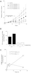
In vivo analysis of FTD accumulation and activity in xenograft models. FTD was administered by daily oral administration or continuous infusion for 14 day to mice subcutaneously implanted with human breast cancer MX-1. (A) Growth curve of xenografts. The tumor volume was measured twice a week and values indicate the means ± SD of the RTV (n=6–7). (B) The amount of FTD incorporated into DNA extracted from MX-1 was measured using HPLC analysis. Tumor for analysis of FTD accumulation was corrected at day 7. The values indicate the means ± SD (n=3). (C) The dUMP levels extracted from MX-1 were also measured using HPLC analysis. FTD was administered by oral administration at 0 h or continuous infusion from 0–24 h. FTD, trifluridine; dUMP, deoxyuridine monophosphate.
Figure 4
Figure 4
Relationship between the antitumor activity of TAS-102 and the amount of FTD incorporated into DNA. The amount of FTD incorporated into DNA in each tumor sample is plotted on the horizontal axis, and the IRs of each antitumor study is plotted on the vertical axis. All data are shown in Table III. A positive correlation was observed between the amount of FTD incorporated into the DNA of tumor cells and the antitumor effect of TAS-102 (Pearson correlation coefficient r=0.92, R2=0.84, P=0.0013). FTD, trifluridine; IRs, inhibition rates.
Figure 5
Figure 5
Antitumor activity of TAS-102 and S-1 and the amount of FTD incorporation into colon cancer xenograft DNA. TAS-102 (150 mg/kg/day) was administered twice daily for 14 days. S-1 (8.3 mg/kg/day) was administered once daily for 14 days. (A) Open circles, triangles and squares indicate control, TAS-102 and S-1 groups, respectively. (B) Amount of FTD incorporated into DNA following TAS-102 treatment. The values are the means ± SD (n=2 to 3). FTD, trifluridine.
Figure 6
Figure 6
TAS-102 prolongs the survival of mice harboring colon cancer xenografts. KM20C colon cancer cells were implanted into the peritoneal cavity of nude mice at a volume of 1×10⁷ cells on day 0. All compounds were administered from day 1 to 28. TAS-102 (150 mg/kg/day) was administered twice daily. S-1 (8.3 mg/kg/day) was administered once daily. Open circles, triangles and squares indicate control, TAS-102 and S-1 groups, respectively.
Figure 7
Figure 7
Ki-67 staining and morphological analysis for MC-2 treated for 14 days with TAS-102. TAS-102 (150 mg/kg/day) was administered to mice harboring MC-2 xenografts twice daily from day 1, and control (A) and TAS-102-treated samples (B) were collected on day 14. Ki-67 staining was performed as described in Materials and methods. Left upper bar indicates 50 μm.

References

    1. Jemal A, Bray F, Center MM, Ferlay J, Ward E, Forman D. Global cancer statistics. CA Cancer J Clin. 2011;61:69–90. - PubMed
    1. Hurwitz H, Fehrenbacher L, Novotny W, et al. Bevacizumab plus irinotecan, fluorouracil, and leucovorin for metastatic colorectal cancer. N Engl J Med. 2004;350:2335–2342. - PubMed
    1. Giantonio BJ, Catalano PJ, Meropol NJ, et al. Bevacizumab in combination with oxaliplatin, fluorouracil, and leucovorin (FOLFOX4) for previously treated metastatic colorectal cancer: results from the Eastern Cooperative Oncology Group Study E3200. J Clin Oncol. 2007;25:1539–1544. - PubMed
    1. Jonker DJ, O’Callaghan CJ, Karapetis CS, et al. Cetuximab for the treatment of colorectal cancer. N Engl J Med. 2007;357:2040–2048. - PubMed
    1. Van Cutsem E, Peeters M, Siena S, et al. Open-label phase III trial of panitumumab plus best supportive care compared with best supportive care alone in patients with chemotherapy-refractory metastatic colorectal cancer. J Clin Oncol. 2007;25:1658–1664. - PubMed

MeSH terms