. 2014 Nov 14;35(43):3013-20, 3020a-3020d.
doi: 10.1093/eurheartj/ehu353. Epub 2014 Sep 17.

Biomechanical factors in atherosclerosis: mechanisms and clinical implications

Affiliations

Biomechanical factors in atherosclerosis: mechanisms and clinical implications

Brenda R Kwak et al. Eur Heart J. .

Abstract

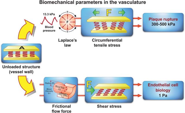
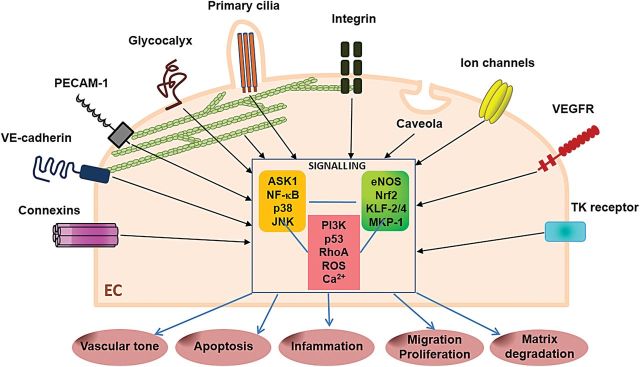
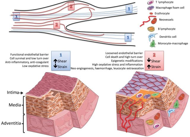
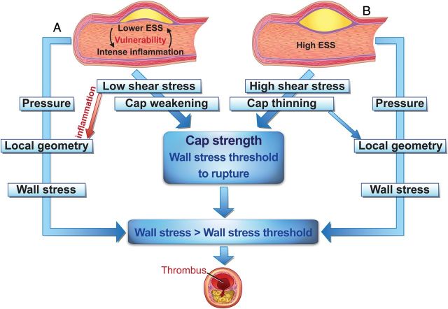
Blood vessels are exposed to multiple mechanical forces that are exerted on the vessel wall (radial, circumferential and longitudinal forces) or on the endothelial surface (shear stress). The stresses and strains experienced by arteries influence the initiation of atherosclerotic lesions, which develop at regions of arteries that are exposed to complex blood flow. In addition, plaque progression and eventually plaque rupture is influenced by a complex interaction between biological and mechanical factors-mechanical forces regulate the cellular and molecular composition of plaques and, conversely, the composition of plaques determines their ability to withstand mechanical load. A deeper understanding of these interactions is essential for designing new therapeutic strategies to prevent lesion development and promote plaque stabilization. Moreover, integrating clinical imaging techniques with finite element modelling techniques allows for detailed examination of local morphological and biomechanical characteristics of atherosclerotic lesions that may be of help in prediction of future events. In this ESC Position Paper on biomechanical factors in atherosclerosis, we summarize the current 'state of the art' on the interface between mechanical forces and atherosclerotic plaque biology and identify potential clinical applications and key questions for future research.

Keywords: Atherosclerosis; Blood flow; Endothelial cell; Haemodynamics; Mechanotransduction; Plaque rupture.


Figures

Figure 1
Figure 1
Biomechanical forces acting on the arterial wall. Blood pressure and blood flow induce forces in the vascular system that deforms the vessel wall. When forces are to be compared, they need to be normalized to area. Force per area is called stress and is expressed in N/m2 or Pascal (Pa). Blood pressure produces a force directed perpendicular to the vessel wall. As a consequence, the cylindrical structure will be stretched circumferentially, resulting in a circumferential stress. Stress in the range of 300–500 kPa is associated with plaque rupture. In contrast, the force induced by a difference in movement of blood and the non-moving vessel wall leads to stress and strain parallel to the surface of endothelial cells. Due to its shearing deformation, this is called a shear stress. This shear stress is of small amplitude (1 Pa) and exerts its main effects through the activation of mechanosensitive receptors and signalling pathways.
Figure 2
Figure 2
Mechanoreceptors and intracellular signalling in arterial endothelium. Schematic representation of a large variety of membrane-associated molecules and microdomains that have been proposed as potential shear stress sensors converting a mechanical signal into a chemical response. Shear stress activates receptor-tyrosine kinase, such as the vascular endothelial growth factor receptor and PECAM-1, which regulate leukocyte adhesion and endothelial cell–endothelial cell coupling as well as mechanoresponsiveness. In addition to these mechanoreceptors, shear stress can also activate ion channels, actin filaments, caveolae, the glycocalyx, primary cilia, and adherence or gap junction proteins. Shear stress influences activation of endothelial cells through multiple mechanisms that target the mitogen-activated protein kinases, nuclear factor-kappa-B, and regulators of these pathways including mitogen-activated protein kinase phosphatase-1, Kruppel-like factors-2 and -4, nuclear factor erythroid 2-related factor, and endothelial nitric oxide synthase.
Figure 3
Figure 3
Effects of shear and strain on the arterial wall. (top) Schematic representation of different biomechanical forces along the arterial tree; 1 = laminar flow (blue lines) imposing a high shear stress parallel to the vascular wall and a low circumferential strain; 2 = arterial regions with a change in the diameter (lack of wall parallelism) and/or proximity to bifurcations (presence of disturbed flow, red line) are subjected to a relatively lower shear stress and higher strain. (bottom left) High shear stress and low strain (‘1’) contribute to maintenance of the physiological properties of the endothelial barrier (anti-coagulant, anti-inflammatory, and anti-oxidant properties) and of the vessel wall (homeostatic cell and matrix turn-over). (bottom right) Low shear and high strain (‘2’) cause endothelial cell death and reduce the physiological endothelial barrier function, thus favouring the formation of atherosclerotic plaques (yellow matter). Plaque progression can also be affected by biomechanical factors inducing an accelerated cell and matrix turn-over, modifications of the vascular stromal cells, inflammation, and intraplaque haemorrhage. This can boost plaque growth and in turn impact on the local flow dynamics, thus generating a vicious circle between biomechanical factors and atherosclerosis.
Figure 4
Figure 4
Concept of the influence of shear stress and wall stress on plaque rupture. Co-localization of peak wall stress and shear stress-induced cap thinning and cap strength will dictate location and timing of plaque rupture. (A) (Excessive) compensatory remodelling induces low shear stress stimulating local inflammation and thereby fibrous cap thinning and plaque weakening, influencing the cap strength, (B) high shear stress induces cap thinning and weakening. Wall stress inside the cap is related to blood pressure and the local cap geometry and thickness. If the local wall stress exceeds the cap strength (the wall stress threshold at which it ruptures), the cap will rupture.

References

    1. Davies PF. Hemodynamic shear stress and the endothelium in cardiovascular pathophysiology. Nat Clin Pract Cardiovasc Med. 2009;6:16–26. - PMC - PubMed
    1. Shaw A, Xu Q. Biomechanical stress-induced signaling in smooth muscle cells: an update. Curr Vasc Pharmacol. 2003;1:41–58. - PubMed
    1. Steinman DA. Simulated pathline visualization of computed periodic blood flow patterns. J Biomech. 2000;33:623–628. - PubMed
    1. Hoefer IE, den Adel B, Daemen MJ. Biomechanical factors as triggers of vascular growth. Cardiovasc Res. 2013;99:276–283. - PubMed
    1. Chouinard-Pelletier G, Jahnsen ED, Jones EA. Increased shear stress inhibits angiogenesis in veins and not arteries during vascular development. Angiogenesis. 2013;16:71–83. - PubMed

Publication types

MeSH terms