Identification and characterization of telocytes in the uterus of the oviduct in the Chinese soft-shelled turtle, Pelodiscus sinensis: TEM evidence
- PMID: 25230849
- PMCID: PMC4302644
- DOI: 10.1111/jcmm.12392
Identification and characterization of telocytes in the uterus of the oviduct in the Chinese soft-shelled turtle, Pelodiscus sinensis: TEM evidence
Abstract
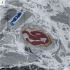
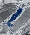
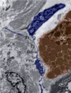
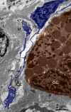
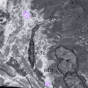
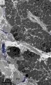
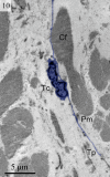
Telocytes (Tcs) are cells with telopodes (Tps), which are very long cellular extensions with alternating thin segments (podomers) and dilated bead-like thick regions known as podoms. Tcs are a distinct category of interstitial cells and have been identified in many mammalian organs including heart, lung and kidney. The present study investigates the existence, ultrastructure, distribution and contacts of Tcs with surrounding cells in the uterus (shell gland) of the oviduct of the Chinese soft-shelled turtle, Pelodiscus sinensis. Samples from the uterine segment of the oviduct were examined by transmission electron microscopy. Tcs were mainly located in the lamina propria beneath the simple columnar epithelium of the uterus and were situated close to nerve endings, capillaries, collagen fibres and secretory glands. The complete morphology of Tcs and Tps was clearly observed and our data confirmed the existence of Tcs in the uterus of the Chinese soft-shelled turtle Pelodiscus sinensis. Our results suggest these cells contribute to the function of the secretory glands and contraction of the uterus.
Keywords: Chinese soft-shelled turtle (Pelodiscus sinensis); telocytes; ultrastructure; uterus.
© 2014 The Authors. Journal of Cellular and Molecular Medicine published by John Wiley & Sons Ltd and Foundation for Cellular and Molecular Medicine.
Figures




Similar articles
-
Telocytes: novel interstitial cells present in the testis parenchyma of the Chinese soft-shelled turtle Pelodiscus sinensis.J Cell Mol Med. 2015 Dec;19(12):2888-99. doi: 10.1111/jcmm.12731. J Cell Mol Med. 2015. PMID: 26769239 Free PMC article.
-
Cellular Evidence of Exosomes in the Reproductive Tract of Chinese Soft-Shelled Turtle Pelodiscus sinensis.J Exp Zool A Ecol Integr Physiol. 2017 Jan;327(1):18-27. doi: 10.1002/jez.2065. Epub 2017 Feb 20. J Exp Zool A Ecol Integr Physiol. 2017. PMID: 28217961
-
Novel cellular evidence of oviduct secretions in the Chinese soft-shelled turtle Pelodiscus sinensis.J Exp Zool A Ecol Genet Physiol. 2015 Nov;323(9):655-65. doi: 10.1002/jez.1957. Epub 2015 Sep 9. J Exp Zool A Ecol Genet Physiol. 2015. PMID: 26350585
-
Uterine Telocytes: A Review of Current Knowledge.Biol Reprod. 2015 Jul;93(1):10. doi: 10.1095/biolreprod.114.125906. Epub 2015 Feb 18. Biol Reprod. 2015. PMID: 25695721 Review.
-
The Cutaneous Telocytes.Adv Exp Med Biol. 2016;913:303-323. doi: 10.1007/978-981-10-1061-3_20. Adv Exp Med Biol. 2016. PMID: 27796896 Review.
Cited by
-
Telocytes in the Normal and Pathological Peripheral Nervous System.Int J Mol Sci. 2020 Jun 17;21(12):4320. doi: 10.3390/ijms21124320. Int J Mol Sci. 2020. PMID: 32560571 Free PMC article. Review.
-
Skin telocytes versus fibroblasts: two distinct dermal cell populations.J Cell Mol Med. 2015 Nov;19(11):2530-9. doi: 10.1111/jcmm.12671. Epub 2015 Sep 28. J Cell Mol Med. 2015. PMID: 26414534 Free PMC article.
-
Ultrastructure damage of oviduct telocytes in rat model of acute salpingitis.J Cell Mol Med. 2015 Jul;19(7):1720-8. doi: 10.1111/jcmm.12548. Epub 2015 Mar 6. J Cell Mol Med. 2015. PMID: 25753567 Free PMC article.
-
Telocytes in the esophageal wall of chickens: a tale of subepithelial telocytes.Poult Sci. 2022 Jul;101(7):101859. doi: 10.1016/j.psj.2022.101859. Epub 2022 Mar 15. Poult Sci. 2022. PMID: 35561461 Free PMC article.
-
Telocytes in gastric lamina propria of the Chinese giant salamander, Andrias davidianus.Sci Rep. 2016 Sep 15;6:33554. doi: 10.1038/srep33554. Sci Rep. 2016. PMID: 27629815 Free PMC article.
References
-
- Faussone-Pellegrini MS, Popescu LM. Telocytes. BioMolecular Concepts. 2011;2:481–9. - PubMed
-
- Rusu MC, Pop F, Hostiuc S, et al. Telocytes form networks in normal cardiac tissues. Histol Histopathol. 2012;27:807–16. - PubMed
-
- Nicolescu MI, Bucur A, Dinca O, et al. Telocytes in parotid glands. Anat Rec (Hoboken) 2012;295:378–85. - PubMed
Publication types
MeSH terms
Substances
LinkOut - more resources
Full Text Sources
Other Literature Sources

