

EWSR1-PBX3: a novel gene fusion in myoepithelial tumors
- PMID: 25231231
- PMCID: PMC4268355
- DOI: 10.1002/gcc.22216
EWSR1-PBX3: a novel gene fusion in myoepithelial tumors
Abstract
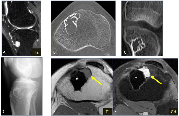
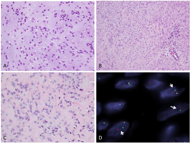
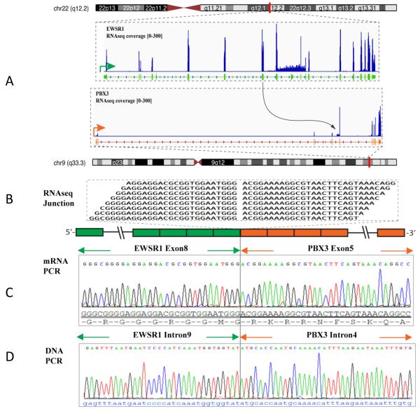
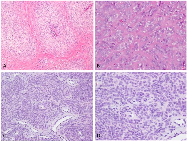
The genetics of myoepithelial tumors (ME) of soft tissue and bone have recently been investigated, with EWSR1-related gene fusions being seen in approximately half of the tumors. The fusion partners of EWSR1 so far described include POU5F1, PBX1, ZNF444 and, in a rare case, ATF1. We investigated by RNA sequencing an index case of EWSR1-rearranged ME of the tibia, lacking a known fusion partner, and identified a novel EWSR1-PBX3 fusion. The fusion was further validated by reverse transcriptase polymerase chain reaction and fluorescence in situ hybridization (FISH). To evaluate if this is a recurrent event, an additional cohort of 22 EWSR1-rearranged ME cases lacking a fusion partner were screened by FISH for abnormalities in PBX3 gene. Thus, two additional cases were identified showing an EWSR1-PBX3 gene fusion. One of them was also intraosseous involving the ankle, while the other occurred in the soft tissue of the index finger. The morphology of the EWSR1-PBX3 fusion positive cases showed similar findings, with nests or sheets of epithelioid to spindle cells in a partially myxoid to collagenous matrix. All three cases showed expression of S100 and EMA by immunohistochemistry. In summary, we report a novel EWSR1-PBX3 gene fusion in a small subset of ME, thereby expanding the spectrum of EWSR1-related gene fusions seen in these tumors. This gene fusion seems to occur preferentially in skeletal ME, with two of the three study cases occurring in intraosseous locations.
© 2014 Wiley Periodicals, Inc.
Figures
References
-
- Alberghini M, Pasquinelli G, Zanella L, Pignatti G, Benini S, Bacchini P, Bertoni F. Primary malignant myoepithelioma of the distal femur. APMIS. 2007;115(4):376–380. - PubMed
-
- Antonescu CR, Zhang L, Chang NE, Pawel BR, Travis W, Katabi N, Edelman M, Rosenberg AE, Nielsen GP, Dal Cin P, Fletcher CD. EWSR1-POU5F1 fusion in soft tissue myoepithelial tumors. A molecular analysis of sixty-six cases, including soft tissue, bone, and visceral lesions, showing common involvement of the EWSR1 gene. Genes Chromosomes Cancer. 2010;49(12):1114–1124. - PMC - PubMed
-
- Brandal P, Panagopoulos I, Bjerkehagen B, Gorunova L, Skjeldal S, Micci F, Heim S. Detection of a t(1;22)(q23;q12) translocation leading to an EWSR1-PBX1 fusion gene in a myoepithelioma. Genes Chromosomes Cancer. 2008;47(7):558–564. - PubMed
-
- Brandal P, Panagopoulos I, Bjerkehagen B, Heim S. t(19;22)(q13;q12) Translocation leading to the novel fusion gene EWSR1-ZNF444 in soft tissue myoepithelial carcinoma. Genes Chromosomes Cancer. 2009;48(12):1051–1056. - PubMed
Publication types
MeSH terms
Substances
Grants and funding
LinkOut - more resources
Full Text Sources
Other Literature Sources
Medical
Molecular Biology Databases
Research Materials
