. 2014 Dec;88(23):13523-46.
doi: 10.1128/JVI.01673-14. Epub 2014 Sep 17.

Elephant endotheliotropic herpesviruses EEHV1A, EEHV1B, and EEHV2 from cases of hemorrhagic disease are highly diverged from other mammalian herpesviruses and may form a new subfamily

Affiliations

Elephant endotheliotropic herpesviruses EEHV1A, EEHV1B, and EEHV2 from cases of hemorrhagic disease are highly diverged from other mammalian herpesviruses and may form a new subfamily

Laura K Richman et al. J Virol. 2014 Dec.

Abstract

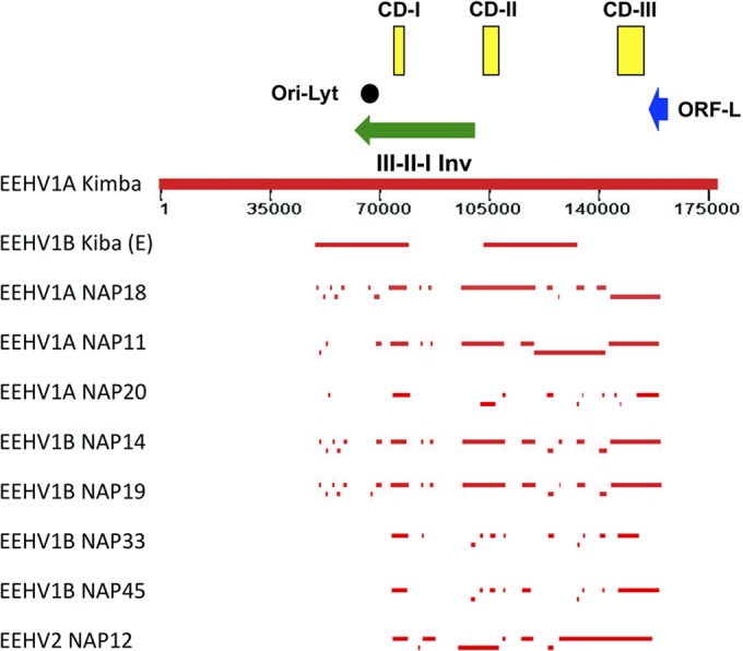
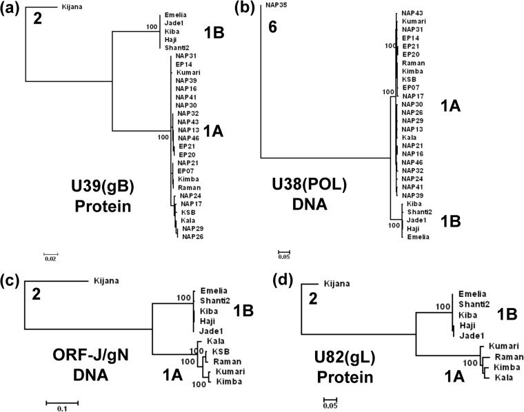
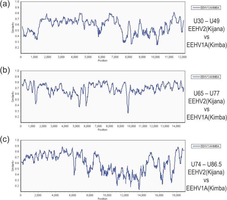
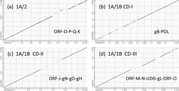
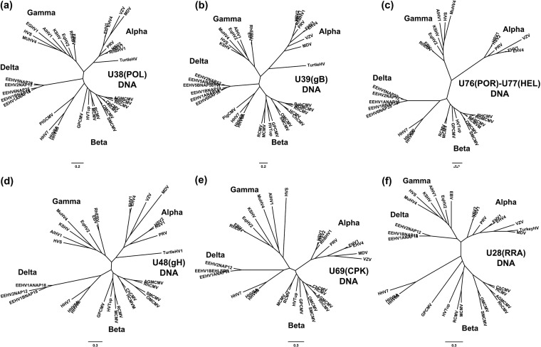
A family of novel endotheliotropic herpesviruses (EEHVs) assigned to the genus Proboscivirus have been identified as the cause of fatal hemorrhagic disease in 70 young Asian elephants worldwide. Although EEHV cannot be grown in cell culture, we have determined a total of 378 kb of viral genomic DNA sequence directly from clinical tissue samples from six lethal cases and two survivors. Overall, the data obtained encompass 57 genes, including orthologues of 32 core genes common to all herpesviruses, 14 genes found in some other herpesviruses, plus 10 novel genes, including a single large putative transcriptional regulatory protein (ORF-L). On the basis of differences in gene content and organization plus phylogenetic analyses of conserved core proteins that have just 20% to 50% or less identity to orthologues in other herpesviruses, we propose that EEHV1A, EEHV1B, and EEHV2 could be considered a new Deltaherpesvirinae subfamily of mammalian herpesviruses that evolved as an intermediate branch between the Betaherpesvirinae and Gammaherpesvirinae. Unlike cytomegaloviruses, EEHV genomes encode ribonucleotide kinase B subunit (RRB), thymidine kinase (TK), and UL9-like origin binding protein (OBP) proteins and have an alphaherpesvirus-like dyad symmetry Ori-Lyt domain. They also differ from all known betaherpesviruses by having a 40-kb large-scale inversion of core gene blocks I, II, and III. EEHV1 and EEHV2 DNA differ uniformly by more than 25%, but EEHV1 clusters into two major subgroups designated EEHV1A and EEHV1B with ancient partially chimeric features. Whereas large segments are nearly identical, three nonadjacent loci totaling 15 kb diverge by between 21 and 37%. One strain of EEHV1B analyzed is interpreted to be a modern partial recombinant with EEHV1A.

Importance: Asian elephants are an endangered species whose survival is under extreme pressure in wild range countries and whose captive breeding populations in zoos are not self-sustaining. In 1999, a novel class of herpesviruses called EEHVs was discovered. These viruses have caused a rapidly lethal hemorrhagic disease in 20% of all captive Asian elephant calves born in zoos in the United States and Europe since 1980. The disease is increasingly being recognized in Asian range countries as well. These viruses cannot be grown in cell culture, but by direct PCR DNA sequence analysis from segments totaling 15 to 30% of the genomes from blood or necropsy tissue from eight different cases, we have determined that they fall into multiple types and chimeric subtypes of a novel Proboscivirus genus, and we propose that they should also be classified as the first examples of a new mammalian herpesvirus subfamily named the Deltaherpesvirinae.


Figures

FIG 1
FIG 1
Schematic map of position coordinates for all sequenced loci of eight EEHV1A, EEHV1B, and EEHV2 genomes compared to the complete EEHV1A(Kimba) genome. This map is drawn to scale with bars representing all of the DNA sequence blocks generated here (Tables 1 and 2) aligned relative to the complete 177,316-kb genome for EEHV1A(Kimba) (24). The data reported by Ehlers et al. (8) for EEHV1B(Kiba) are also given for comparison in the top line. The locations of the predicted Ori-Lyt dyad symmetry locus (black circle), the large 40-kb inverted (Inv) core domain I, II, and III segment (green arrow), the putative immediate early-like ORF-L transactivator protein coding region (blue arrow), and the three major hypervariable domains CD-I, CD-II, and CD-III (yellow boxes) are all indicated.
FIG 2
FIG 2
Linear phylogenetic trees illustrating the EEHV1A versus EEHV1B diaspora among multiple strains within the major chimeric domains. Maximum likelihood phylogenetic dendrograms for two sets of proteins and two DNA loci derived from multiple independent EEHV1 strains. The diagrams include equivalent data from the orthologous prototype genes of either EEHV2 or EEHV6 as outgroups. (a) Set of 28 examples of the EEHV1 U39(gB) protein mapping within CD-I (EEHV1A [1A] [851 aa], EEHV1B [1B] [847 aa], and EEHV2 [2] [851 aa]). (b) Set of 28 examples of the EEHV1 U38(POL) gene DNA locus mapping within CD-I (1A [2,319 bp], 1B [2,304 bp], and 2 [2,316 bp]). (c) Set of 10 examples of the combined EEHV1 U45.7(ORF-J) plus U46(ORF-N) gene DNA locus mapping within CD-II (1A [753 bp], 1B [700 bp], and 2 [780 bp]). (d) Set of 9 examples of the EEHV1 U82(ORF-L) protein mapping within CD-III (1A [263 aa], 1B [265 aa], and 2 [266 aa]). After gaps were omitted, the final data sets totalled 843 aa in panel a, 2301 bp in panel b, 666 bp in panel c, and 261 aa in panel d. PCR data given here for ORF-J/gN of EEHV1A(NAP23, Kimba) and for gL of EEHV1B(EP18, Emelia) match those from their complete genomes (GenBank accession nos. KC618527 and KC462164). Data for EEHV1A(Raman) derived from GenBank accession no. KC462165 was included in all four panels for comparison. Bootstrap values (100 replicates), distance scales (number of substitutions per site).
FIG 3
FIG 3
Divergence patterns across the three largest sequenced segments of EEHV2 compared to EEHV1A. The diagrams show SimPlot comparisons of the nucleotide identity patterns for each of the three largest sequenced segments of EEHV2(NAP12) versus their matching orthologous regions in EEHV1A(NAP23, Kimba) (GenBank accession no. KC618527) (24). (a) U30-to-U49 segment. This segment is 12.3 kb and encompasses EEHV2(NAP12) genes U30 to U49, map coordinates 93,790 to 106,934. (b) U65-to-U77 segment. This segment is 14.8 kb and encompasses U65 to U77, map coordinates 127,341 to 141,906. (c) U74-to-U86.5 segment. This segment is 17.5 kb and encompasses U74 to U82, map coordinates 137,703 to 156,114. The relevant source GenBank DNA file accession numbers are included in Table S1 in the supplemental material.
FIG 4
FIG 4
Dot matrix alignments encompassing major areas of divergence for EEHV2 or EEHV1B versus EEHV1A. Examples of DNA level dot matrix alignments produced in BLAST2.2.22 as available online at NCBI. (a) Highly divergent ORF-O-P-K locus in EEHV2. The alignment from U74(PAF) to U86.5(ORF-L) was created by comparing EEHV2(NAP12) positions 1 to 17,536 from GenBank accession no. KC609754 with EEHV1A(NAP23, Kimba) positions 137,7000 to 156,117 from GenBank accession no. KC618527. Match-mismatch settings were 2,3, and gapcode was 2,2. The four mismatch gaps encompass parts of ORF-O, -P, -Q, and -K in EEHV1A, with the displaced segment resulting from the absence of ORF-Q in EEHV2. (b) CD-I locus in EEHV1B. Comparison of U39(gB) and U38(POL) using GenBank accession no. HM568523 (positions 1 to 5,947) from EEHV1A(NAP18, Kala) with GenBank accession no. HM568550 (positions 1 to 5,899) from EEHV1B(NAP19, Haji). Match-mismatch settings were 1,4, and gapcode was 5,2. (c) CD-II locus in EEHV1B. Comparison of U29(TRI1) to U51(vGPCR1) using GenBank accession no. HM568525 (positions 1 to 13,593) from EEHV1A(NAP18, Kala) with GenBank accession no. HM568538 (positions 1 to 13,593) from EEHV1B(NAP14, Kiba). Match-mismatch settings were 1,−3, and gapcode was 2,2. The central 3.2-kb gap represents part of U45.7(ORF-J), plus all of U46(gN), U47(gO), and U48(gH). (d) CD-III locus in EEHV1B (left-hand side [LHS]). Comparison of U77(HEL) to U85.5(ORF-O) using GenBank accession no. JX011082 (positions 223 to 4,279) from EEHV1B(NAP14, Kiba) with GenBank accession no. KC854707 (positions 1 to 4,293) from EEHV1A(NAP18, Kala). Match-mismatch settings were 1,−2, and gapcode was linear. The small displacement at the first gap represents a 220-bp deletion within the ORF-N(vCXCL1) gene of EEHV1B.
FIG 5
FIG 5
EEHV1A-B chimeric domain I and II patterns and boundaries relative to EEHV2. The diagrams show SimPlot comparisons of the nucleotide identity patterns between EEHV1A, EEHV1B, and EEHV2 across the first two EEHV1B chimeric domains CD-I (3.0 kb) encompassing glycoprotein B and POL(N-term) (N-term stands for N terminus) and CD-II (3.7 kb) encompassing ORF-J(C-term)-gN-gO-gH and TK(C-term) (C-term stands for C terminus). Vertical arrows indicate positions of chimeric domain boundaries. (a) CD-I. The 5,000-bp U39(gB)-U38(POL) segment from EEHV1A(Kala, NAP18) is compared with EEHV1B(Haji, NAP19) (green) and with EEHV2(Kijana, NAP12) (gray), map coordinates 73,959 to 79,043. (b) CD-II. The 6,000-bp U27(PPF)-U48.5(TK) segment from EEHV1A(Kala, NAP18) is compared with EEHV1B(Kiba, NAP14) (red) and with EEHV2(Kijana, NAP12) (gray), map coordinates 100,981 to 106,909. (c) CD-II. The 6,000-bp U27(PPF)-U48.5(TK) segment from EEHV1A(Kala, NAP18) is compared with EEHV1B(Haji, NAP19) (green) and with EEHV2(Kijana, NAP12) (gray), map coordinates 100,981 to 106,909. The relevant source DNA file accession numbers are included in Table S1 in the supplemental material.
FIG 6
FIG 6
EEHV1A-1B chimeric domain III patterns and boundaries relative to EEHV2. The diagrams show SimPlot comparisons of the nucleotide identity patterns between EEHV1A, EEHV1B, and EEHV2 across both sides of EEHV1B chimeric domain CD-III (8.3-kb) encompassing ORF-M–ORF-N(vCXCL1)–UDG–gL–ORF-O–ORF-P–ORF-Q. (a) CD-III left-hand side (LHS) from U77(HEL, C-term) to U82.5(ORF-O, C-term). A 4,330-bp segment of EEHV1A(Kala, NAP18), is compared with EEHV1B(Kiba, NAP14) (red) and EEHV2(Kijana, NAP12) (gray), map coordinates 143,625 to 147,955. (b) CD-III left-hand side from U77(HEL, C-term) to U82.5(ORF-O, C-term). A 4,330-bp segment of EEHV1A(Kala, NAP18) is compared with EEHV1B(Haji, NAP19) (green) and with EEHV2(Kijana, NAP12) (gray), map coordinates 143,625 to 147,955. (c) CD-III right-hand side (RHS) from U82.5(ORF-O, C-term) to U85.5(ORF-K, N-term). A 4,750-bp segment of EEHV1A(Kumari, NAP11), is compared with a matching 4,200-bp segment of EEHV1B(Kiba, NAP14) (red) and a 4,650-bp segment of EEHV2(Kijana, NAP12) (gray), map coordinates 143,401 to 153,115. (d) CD-III right-hand side from U83.5(ORF-O, C-term) to U85.5(ORF-K N-term). The same segment of EEHV1A(Kumari) is compared with EEHV1B(Haji, NAP19) (green) and EEHV2(Kijana, NAP12) (gray), map coordinates 148,401 to 153,115. The relevant GenBank file accession numbers are included in Table S1 in the supplemental material.
FIG 7
FIG 7
Radial phylogenetic trees for six highly conserved core EEHV proteins. The diagrams present radial amino acid dendrograms generated by Bayesian maximum likelihood in MEGA5 that compare several of the most conserved core proteins encoded by EEHV genomes (proposed Deltaherpesvirinae [Delta]) with orthologues found in all other mammalian herpesvirus subfamilies. Each locus includes data for up to five EEHV species, including the EEHV1A, EEHV1B, and EEHV2 proteins analyzed in this paper, together with any available data for the prototype EEHV5 and EEHV6 versions from the accompanying paper by Zong et al. (26). The EEHV proteins are compared with matching segments of selected orthologues from up to 32 other representative herpesviruses from the three currently named mammalian subfamilies: Alphaherpesvirinae (Alpha), Gammaherpesvirinae (Gamma), and Betaherpesvirinae i(Beta), including the Roseolovirus species HHV6A, HHV6B, and HHV7 and pig CMV. Alternative or more-formal virus names as well as GenBank accession numbers for the relevant non-EEHV proteins used are listed in the supplemental material. The final segment sizes evaluated were as follows for panels a to f: (a) U38(POL), DNA polymerase, 668 aa; (b) U39(gB), glycoprotein B, 700 aa; (c) U48(gH), glycoprotein H, 497 aa; (d) U72(gM), glycoprotein M, 329 aa; (e) U57(MCP), major capsid protein, 643 aa; (f) U69(CPK), conserved protein kinase, 285 aa. Bootstrap values for the linear forms of these types of trees are presented in Zong et al. (26).
FIG 8
FIG 8
Radial phylogenetic trees for six poorly conserved EEHV proteins. Similar to Fig. 7 except that the six additional proteins evaluated were selected from among the least conserved proteins. Final segment sizes evaluated were as follows for panels a to f: (a) U48.5(TK), thymidine kinase, 238 aa; (b) U73(OBP), origin binding protein, 256 aa; (c) U27.5(RRB), ribonucleotide kinase B subunit, 175 aa; (d) U28(RRA), ribonucleotide reductase A subunit, 575 aa; (e) U27(PPF, ORF-I), polymerase processivity factor, 241 aa; (f) U51(vGPCR1), G-protein-coupled receptor 1, 171 aa. The CCR3 protein from Loxodonta africana (LOXCCR3, GenBank accession no. XP_003409912) was included for comparison in panel f.
FIG 9
FIG 9
DNA level radial phylogenetic trees for six selected EEHV gene loci. Similar to Fig. 7 and 8 except that the six gene loci chosen were evaluated at the DNA level, not the protein level. The final segment sizes involved were as follows for panels a to f: (a) U38(POL) locus, DNA polymerase, DPOL, 1,668 bp; (b) U39(gB) locus, glycoprotein B, 932 bp; (c) U76(POR)-U77(HEL) locus, portal plus helicase subunit, 1,457 bp; (d) U48(gH) locus, glycoprotein H, 887 bp; (e) U69(CPK) locus, conserved protein kinase, 578 bp; and (f) U28/RRA locus, 1,226 bp. The DNA POL protein from elephant gammaherpesvirus 1 (EGHV1) (GenBank accession no. EU085379) is also included for comparison in panel a.

Comment in

  • Trunkloads of viruses.
    Pellett PE. Pellett PE. J Virol. 2014 Dec;88(23):13520-2. doi: 10.1128/JVI.02359-14. Epub 2014 Sep 17. J Virol. 2014. PMID: 25231304 Free PMC article.

Similar articles

Cited by

References

    1. Jacobson ER, Sundberg JP, Gaskin JM, Kollias GV, O'Banion MK. 1986. Cutaneous papillomas associated with a herpesvirus-like infection in a herd of captive African elephants. J. Am. Vet. Med. Assoc. 189:1075–1078. - PubMed
    1. McCully RM, Basson PA, Pienaar JG, Erasmus BJ, Young E. 1971. Herpes nodules in the lung of the African elephant (Loxodonta africana (Blumebach, 1792)). Onderstepoort J. Vet. Res. 38:225–235. - PubMed
    1. Ossent P, Guscetti F, Metzler AE, Lang EM, Rübel A, Hauser B. 1990. Acute and fatal herpesvirus infection in a young Asian elephant (Elephas maximus). Vet. Pathol. 27:131–133. 10.1177/030098589002700212. - DOI - PubMed
    1. Richman LK, Montali RJ, Garber RL, Kennedy MA, Lehnhardt J, Hildebrandt T, Schmitt D, Hardy D, Alcendor DJ, Hayward GS. 1999. Novel endotheliotropic herpesviruses fatal for Asian and African elephants. Science 283:1171–1176. 10.1126/science.283.5405.1171. - DOI - PubMed
    1. Hayward GS. 2012. Conservation: clarifying the risk from herpesvirus to captive Asian elephants. Vet. Rec. 170:202–203. 10.1136/vr.e1212. - DOI - PMC - PubMed

Publication types

Associated data

LinkOut - more resources