. 2014 Dec;88(23):13747-58.
doi: 10.1128/JVI.02040-14. Epub 2014 Sep 17.

Characterization of an alphamesonivirus 3C-like protease defines a special group of nidovirus main proteases

Affiliations

Characterization of an alphamesonivirus 3C-like protease defines a special group of nidovirus main proteases

Sandra Blanck et al. J Virol. 2014 Dec.

Abstract

Cavally virus (CavV) and related viruses in the family Mesoniviridae diverged profoundly from other nidovirus lineages but largely retained the characteristic set of replicative enzymes conserved in the Coronaviridae and Roniviridae. The expression of these enzymes in virus-infected cells requires the extensive proteolytic processing of two large replicase polyproteins, pp1a and pp1ab, by the viral 3C-like protease (3CL(pro)). Here, we show that CavV 3CL(pro) autoproteolytic cleavage occurs at two N-terminal (N1 and N2) and one C-terminal (C1) processing site(s). The mature form of 3CL(pro) was revealed to be a 314-residue protein produced by cleavage at FKNK1386|SAAS (N2) and YYNQ1700|SATI (C1). Site-directed mutagenesis data suggest that the mesonivirus 3CL(pro) employs a catalytic Cys-His dyad comprised of CavV pp1a/pp1ab residues Cys-1539 and His-1434. The study further suggests that mesonivirus 3CL(pro) substrate specificities differ from those of related nidovirus proteases. The presence of Gln (or Glu) at the P1 position was not required for cleavage, although residues that control Gln/Glu specificity in related viral proteases are retained in the CavV 3CL(pro) sequence. Asn at the P2 position was identified as a key determinant for mesonivirus 3CL(pro) substrate specificity. Other positions, including P4 and P1', each are occupied by structurally related amino acids, indicating a supportive role in substrate binding. Together, the data identify a new subgroup of nidovirus main proteases and support previous conclusions on phylogenetic relationships between the main nidovirus lineages.

Importance: Mesoniviruses have been suggested to provide an evolutionary link between nidovirus lineages with small (13 to 16 kb) and large (26 to 32 kb) RNA genome sizes, and it has been proposed that a specific set of enzymes, including a proofreading exoribonuclease and other replicase gene-encoded proteins, play a key role in the major genome expansion leading to the currently known lineages of large nidoviruses. Despite their smaller genome size (20 kb), mesoniviruses retained most of the replicative domains conserved in large nidoviruses; thus, they are considered interesting models for studying possible key events in the evolution of RNA genomes of exceptional size and complexity. Our study provides the first characterization of a mesonivirus replicase gene-encoded nonstructural protein. The data confirm and extend previous phylogenetic studies of mesoniviruses and related viruses and pave the way for studies into the formation of the mesonivirus replication complex and functional and structural studies of its functional subunits.


Figures

FIG 1
FIG 1
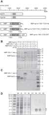
Autocatalytic release of the putative CavV 3CLpro domain from flanking sequences and determination of the C-terminal 3CLpro autoprocessing site. (A) Schematic representation of CavV pp1a/1ab replicase polyproteins and fusion protein constructs used in this experiment. The putative 3CLpro domain is indicated in dark gray, and transmembrane domains are shown in light gray. 3CLpro mut, putative 3CLpro domain containing a Cys-1539-to-Ala change in the expressed sequence. (B and C) Expression analysis of the MBP-pp1a-1343-1720-His6 (wt) and MBP-pp1a-1343-1720_C1539A-His6 (mut) fusion proteins. Expression was induced with 1 mM IPTG for 4 h at 18°C. Cell lysates obtained from IPTG-induced and noninduced cells were analyzed in a 14% SDS-polyacrylamide gel (B) or by Western blotting using an MBP-specific monoclonal antibody. The products of the expression control pMAL-c2 (MBP-lacZα) and the mutant form of the MBP-3CLpro fusion protein (3CLpro mut) are indicated to the left. Two putative processing products derived from the MBP-3CLpro wt construct are indicated by filled circles. Molecular masses (in kDa) of prestained marker proteins are indicated to the right. (D) Purification of C-terminal (GST-containing) cleavage products produced by 3CLpro-mediated cleavage of the MBP-pp1a-1343-1720-GST fusion protein by glutathione Sepharose chromatography. GST-containing cleavage products eluted in fraction 4 (F4) were subjected to N-terminal sequence analysis by Edman degradation. All three cleavage products were shown to have the N terminus Ser-Ala-Thr-…, suggesting that cleavage occurred at 1700Q|S1701 in the CavV pp1a/pp1ab sequence. Molecular masses (in kDa) of marker proteins are indicated to the left. SF, soluble protein fraction; IF, insoluble fraction; FT, column flowthrough fraction; W, wash fraction; F3 to F6, elution fractions 3 to 6.
FIG 2
FIG 2
Determination of the N-terminal 3CLpro autoprocessing site. (A) Schematic representation of the protein construct used in this experiment. The construct lacked the C-terminal 3CLpro residue (Gln-1700) and contained a short C-terminal linker sequence, including a heptahistidine (His7) tag. (B) Purification by Ni-IMAC of C-terminally His7-tagged processing products derived from 3CLpro-mediated cleavage of the fusion protein construct. N-terminal sequence analysis of the protein present in fraction 22 revealed the sequence Ser-Ala-Ala-…, suggesting that cleavage occurred at 1386K|S1387 in the CavV pp1a/pp1ab sequence. Molecular masses (in kDa) of marker proteins are indicated to the left. SF, soluble protein fraction; IF, insoluble protein fraction; FT, column flowthrough fraction; W, wash fraction; 16 to 24, elution fractions.
FIG 3
FIG 3
Mutation analysis of N- and C-terminal CavV 3CLpro autoprocessing sites. (A) Schematic representation of the fusion protein constructs used to study effects of amino acid substitutions at one of the N-terminal (N1 and N2) or the C-terminal (C1) cleavage site. Positions and sequences of individual cleavage sites are indicated, and calculated molecular masses of processing products are given. TM, transmembrane domains. (B and C) Expression and proteolytic processing of MBP-pp1a-1343-1720-GST-derived proteins containing single or multiple Ala substitutions for residues at the 3CLpro N1 (B), N2 (B), and C1 (C) autoprocessing sites, respectively. Substitutions introduced in the respective protein constructs are given above each lane. As controls, MBP-1343-1720-GST wt, MBP-pp1a-1343-1720_C1539A-GST mut, and MBP-lacZα were used. Total cell lysates obtained after induction of expression for 4 h at 18°C were analyzed in a 14% SDS-polyacrylamide gel. Recombinantly expressed N1-C1 and N2-C1 proteins were used as marker proteins. 3CLpro N1-C1, pp1a-1377-1700_H1434A_C1539A; 3CLpro N2-C1, pp1a-1387-1700_H1434A_C1539A. Fusion proteins and processing products were detected by Western blotting using GST-specific (bottom) and CavV 3CLpro-specific (top) antibodies, respectively. The sizes (in kDa) of marker proteins are indicated to the right. The position of the fully processed form of the 3CLpro, which comigrates with the recombinant 3CLpro N2-C1 protein, is indicated by an arrowhead to the left in B and C.
FIG 4
FIG 4
Identification of the 3CLpro in CavV-infected cells. C6/36 cells were mock infected (lane 1) or infected with CavV (lane 2). Western blot analysis of total protein extracts obtained at 48 h p.i. using CavV 3CLpro-specific antiserum is shown. Molecular masses (in kDa) of prestained size markers (M) are indicated to the left. As additional size markers, two recombinant forms representing differentially processed forms of the 3CLpro domain (described in the legend to Fig. 3) were used. 3CLpro N1-C1, pp1a-1377-1700_C1539A_H1434A (lane 3); 3CLpro N2-C1, pp1a-1387-1700_C1539A_H1434A (lane 4).
FIG 5
FIG 5
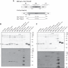
Mutation analysis of putative catalytic and substrate-binding residues. (A and B) pMAL-c2-[pp1a-1343-1720-GST] plasmid DNA was used to express CavV pp1a/pp1ab residues 1343 to 1720 in E. coli TB1 cells. Protein expression was induced with 1 mM IPTG (right) for 4 h at 18°C or not induced (left), and total cell lysates were analyzed by SDS-PAGE. (A) Coomassie blue-stained 14% SDS-polyacrylamide gel. (B) Western blot analysis using CavV 3CLpro-specific rabbit antiserum. The MBP-pp1a-1343-1720-GST fusion contained either the 3CLpro wild-type sequence or the same sequence with Ala substitutions for single-amino-acid residues potentially involved in catalysis (Cys-1539, Asp-1465, His-1434, Asp-1467, Glu-1460, and Glu-1470) or specific binding of the P1 residue (His-1554 and Thr-1534). (B) The unprocessed fusion protein precursor (MBP-pp1a-1343-1720-GST), the N-terminally processed cleavage product (3CLpro-pp1a-1700-1720-GST), and the fully processed 3CLpro domain (3CLpro) are indicated to the right. Molecular masses (in kDa) of marker proteins are indicated to the left.
FIG 6
FIG 6
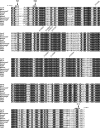
Sequence alignment of mesonivirus 3CLpro domains. Shown are the putative 3CLpro domains of representative mesoniviruses, together with the predicted N- and C-terminal processing sites (indicated as N1, N2, and C1, respectively; see the text for details). Amino acid numbering is according to the position in the CavV pp1a/pp1ab amino acid sequence. Ala substitutions for specific residues in CavV 3CLpro that were characterized in this study are indicated. Filled diamonds denote residues near the scissile bond of the N1, N2, and C1 cleavage site, respectively, that were characterized by site-directed mutagenesis in this study. Abbreviations of virus names are the following: CavV, Cavally virus (isolate C79); NDiV, Nam Dinh virus (isolate 02VN178); HoustonV, Houston virus (strain V3982); HanaV, Hana virus (strain A4/CI/2004); NséV, Nsé virus (strain F24/CI/2004); MénoV, Méno virus (strain E9/CI/2004); NgewotanV, Ngewotan virus (strain JKT9982); BontangV, Bontang virus (strain JKT7774); KSaV, Karang Sari virus (strain JKT10701); KPhV, Kamphang Phet virus (strain KP84-0344); CASV, Casuarina virus (isolate 0071).
FIG 7
FIG 7
3CLpro autoprocessing sites in mesoni-, arteri-, and coronaviruses. Alignments of the P5-P5′ positions of N- and C-terminal autoprocessing sites of nidovirus 3CLpro domains were used to produce sequence logo presentations (43). Alignments included sequences from 11 mesoniviruses (Fig. 6 and the legend to Fig. 6), 5 arteriviruses (from 4 approved species) (44), and 20 coronaviruses, the latter representing all 17 approved alpha-, beta-, and gammacoronavirus species and another 3 yet-to-be-approved species from the genus Deltacoronavirus (4, 45). The height of each letter (amino acid residue) is proportional to the frequency of a specific residue at a given position. Strictly conserved residues at the P1 and P2 positions are indicated in gray. Accession numbers of arterivirus and coronavirus sequences used in this analysis include the following: equine arteritis virus (Bucyrus), NC_002532; simian hemorrhagic fever virus, NC_003092; porcine reproductive and respiratory syndrome virus (PRRSV) (VR-2332), U87392; PRRSV (Lelystad), M96262; lactate dehydrogenase elevating virus (Plagemann), NC_001639; transmissible gastroenteritis coronavirus (Purdue), AJ271965; human coronavirus (HCoV) 229E, AF304460; HCoV-NL63 (Amsterdam 1), AY567487; Miniopterus bat CoV 1 (Mi-BatCoV 1), EU420138; Mi-BatCoV HKU8, EU420139; porcine epidemic diarrhea virus, AF353511; Rhinolophus bat CoV, EF203065; Scotophilus bat CoV 512, DQ648858; bovine coronavirus (Mebus), U00735; HCoV-HKU1, AY597011; mouse hepatitis virus (JHM), AC_000192; Pipistrellus bat CoV HKU5 (Pi-BatCoV HKU5), EF065509; Rousettus bat CoV HKU9 (Ro-BatCoV HKU9), EF065513; severe acute respiratory syndrome-CoV (Urbani), AY278741; Tylonycteris bat CoV HKU4 (Ty-BatCoV HKU4), EF065505; infectious bronchitis virus (Beaudette), M95169; beluga whale CoV SW1, EU111742; munia CoV HKU13, FJ376622; bulbul CoV HKU11, FJ376619; thrush CoV HKU12, FJ376621.

Similar articles

Cited by

References

    1. de Groot RJ, Cowley JA, Enjuanes L, Faaberg KS, Perlman S, Rottier PJM, Snijder EJ, Ziebuhr J, Gorbalenya AE. 2012. Order Nidovirales, p 785–795 In King AMQ, Adams MJ, Carstens EB, Lefkowitz EJ. (ed), Virus taxonomy. Elsevier, Amsterdam, the Netherlands.
    1. Lauber C, Ziebuhr J, Junglen S, Drosten C, Zirkel F, Nga PT, Morita K, Snijder EJ, Gorbalenya AE. 2012. Mesoniviridae: a proposed new family in the order Nidovirales formed by a single species of mosquito-borne viruses. Arch. Virol. 157:1623–1628. 10.1007/s00705-012-1295-x. - DOI - PMC - PubMed
    1. Cowley JA, Dimmock CM, Spann KM, Walker PJ. 2000. Gill-associated virus of Penaeus monodon prawns: an invertebrate virus with ORF1a and ORF1b genes related to arteri- and coronaviruses. J. Gen. Virol. 81:1473–1484. - PubMed
    1. de Groot RJ, Baker SC, Baric R, Enjuanes L, Gorbalenya AE, Holmes KV, Perlman S, Poon L, Rottier PJM, Talbot PJ, Woo PCY, Ziebuhr J. 2012. Family Coronaviridae, p 806–828 In King AMQ, Adams MJ, Carstens EB, Lefkowitz EJ. (ed), Virus taxonomy. Elsevier, Amsterdam, the Netherlands.
    1. Graham RL, Donaldson EF, Baric RS. 2013. A decade after SARS: strategies for controlling emerging coronaviruses. Nat. Rev. Microbiol. 11:836–848. 10.1038/nrmicro3143. - DOI - PMC - PubMed

Publication types

LinkOut - more resources