. 2014 Dec;88(23):13709-21.
doi: 10.1128/JVI.01732-14. Epub 2014 Sep 17.

Analysis of the evolution and structure of a complex intrahost viral population in chronic hepatitis C virus mapped by ultradeep pyrosequencing

Affiliations

Analysis of the evolution and structure of a complex intrahost viral population in chronic hepatitis C virus mapped by ultradeep pyrosequencing

Brendan A Palmer et al. J Virol. 2014 Dec.

Abstract

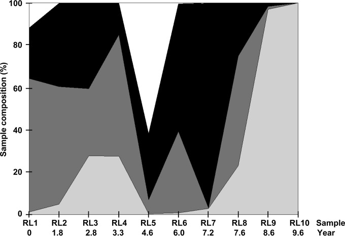
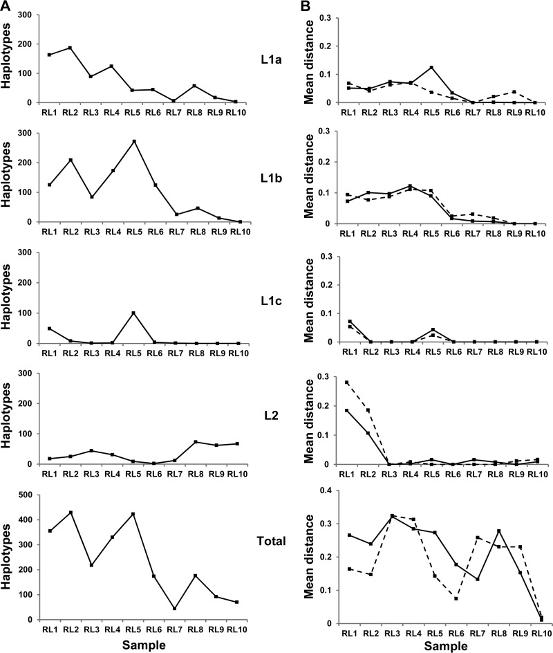
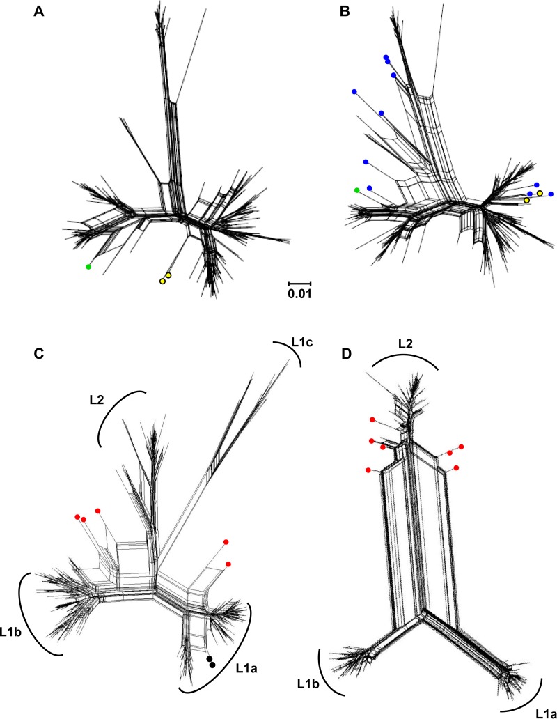
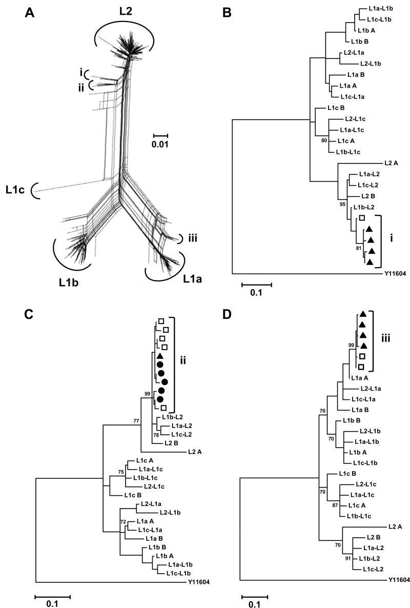
Hepatitis C virus (HCV) causes chronic infection in up to 50% to 80% of infected individuals. Hypervariable region 1 (HVR1) variability is frequently studied to gain an insight into the mechanisms of HCV adaptation during chronic infection, but the changes to and persistence of HCV subpopulations during intrahost evolution are poorly understood. In this study, we used ultradeep pyrosequencing (UDPS) to map the viral heterogeneity of a single patient over 9.6 years of chronic HCV genotype 4a infection. Informed error correction of the raw UDPS data was performed using a temporally matched clonal data set. The resultant data set reported the detection of low-frequency recombinants throughout the study period, implying that recombination is an active mechanism through which HCV can explore novel sequence space. The data indicate that polyvirus infection of hepatocytes has occurred but that the fitness quotients of recombinant daughter virions are too low for the daughter virions to compete against the parental genomes. The subpopulations of parental genomes contributing to the recombination events highlighted a dynamic virome where subpopulations of variants are in competition. In addition, we provide direct evidence that demonstrates the growth of subdominant populations to dominance in the absence of a detectable humoral response.

Importance: Analysis of ultradeep pyrosequencing data sets derived from virus amplicons frequently relies on software tools that are not optimized for amplicon analysis, assume random incorporation of sequencing errors, and are focused on achieving higher specificity at the expense of sensitivity. Such analysis is further complicated by the presence of hypervariable regions. In this study, we made use of a temporally matched reference sequence data set to inform error correction algorithms. Using this methodology, we were able to (i) detect multiple instances of hepatitis C virus intrasubtype recombination at the E1/E2 junction (a phenomenon rarely reported in the literature) and (ii) interrogate the longitudinal quasispecies complexity of the virome. Parallel to the UDPS, isolation of IgG-bound virions was found to coincide with the collapse of specific viral subpopulations.


Figures

FIG 1
FIG 1
The area graph represents the percentages of overall sequences contributing to the subpopulations of L1a (dark gray), L1b (black), L1c (white), and L2 (light gray) per sample over time.
FIG 2
FIG 2
(A) Number of unique full-length nucleotide haplotypes for each group (L1a, L1b, L1c, and L2 and the combined total) recovered per time point. (B) dN (solid line) and dS (dashed line) measurements across HVR1 are given for those nucleotide sequences with frequencies of >0.001. HVR1 is defined between position 196 and position 276 or 279 of the amplicon sequence. The numbers of (non)synonymous substitutions per (non)synonymous site from averaging over all sequence pairs within each group per time point are shown. The importance of separating haplotypes into defined groups is illustrated by the deceptively high dN and dS values recovered when the elements in the data set were not segregated.
FIG 3
FIG 3
Phylogenetic networks generated by NNet. (A) Complete clonal data set and (B) clonal data set supplemented with 12 in silico-generated interlineage recombinants (blue circles). Yellow circles denote recombinants FJ744095 and JQ743309 identified previously through clonal analysis, while the green circle denotes the novel HM363402 clonal recombinant identified in this study. (C) UDPS data subsets RL1 and (D) RL8 are given as representative images for samples containing a complex population and a less-diverse population of haplotypes (355 and 176 unique nucleotide sequences, respectively). Red circles identify inter(sub)lineage recombinants. Black circles identify putative intralineage recombinants. The genetic distance of 0.01 nucleotide substitutions per site is given by the scale bar.
FIG 4
FIG 4
Intersample maintenance of recombinants. (A) Composite phylogenetic network of the RL7-to-RL10 subset. Haplotype clusters displaying split decomposition signals are identified (i to iii). (B to D) Phylogenetic analysis of clusters i to iii, consensus sequences of the (sub)lineages, and interlineage recombinant sequences generated in silico using a rooted general time-reversible model (GTR + G + I). The data demonstrate intersample maintenance and evolution of recombinant haplotypes arguing in favor of the hypothesis of the recombinant sequences arising in situ within the host. Sequences are identified by time points as black circles (RL7), open squares (RL8), and black triangles (RL9). Sequences arising from RL10 did not feature in groups i to iii. A genotype 4a strain (Y11604) was used as the reference outgroup. Bootstrap values (of 1,000 resamplings) above 70 are shown. The genetic distance of 0.01 nucleotide substitutions per site is given by the scale bar.
FIG 5
FIG 5
Detection of IgG-bound virus within the UDPS data set. Five of nine unique E2 amino acid sequences translated from RNA obtained following albumin/IgG column extraction of virions were present within the UDPS data set. The percentages (sample proportion) of these sequences at the nucleotide (gray lines) and amino acid (black lines) levels within the overall sample population are given. The respective sequence accession numbers are GQ985332 (A), GQ985336 (B), GQ985372 (C), HM363384 (D), and GQ985333 (E). Black arrows indicate the sampling point.

Similar articles

Cited by

References

    1. Yamane D, McGivern DR, Masaki T, Lemon SM. 2013. Liver injury and disease pathogenesis in chronic hepatitis C. Curr. Top. Microbiol. Immunol. 369:263–288. 10.1007/978-3-642-27340-7_11. - DOI - PubMed
    1. Deleersnyder V, Pillez A, Wychowski C, Blight K, Xu J, Hahn YS, Rice CM, Dubuisson J. 1997. Formation of native hepatitis C virus glycoprotein complexes. J. Virol. 71:697–704. - PMC - PubMed
    1. Penin F, Combet C, Germanidis G, Frainais PO, Deleage G, Pawlotsky JM. 2001. Conservation of the conformation and positive charges of hepatitis C virus E2 envelope glycoprotein hypervariable region 1 points to a role in cell attachment. J. Virol. 75:5703–5710. 10.1128/JVI.75.12.5703-5710.2001. - DOI - PMC - PubMed
    1. von Hahn T, Yoon JC, Alter H, Rice CM, Rehermann B, Balfe P, McKeating JA. 2007. Hepatitis C virus continuously escapes from neutralizing antibody and T-cell responses during chronic infection in vivo. Gastroenterology 132:667–678. 10.1053/j.gastro.2006.12.008. - DOI - PubMed
    1. Wang GP, Sherrill-Mix SA, Chang KM, Quince C, Bushman FD. 2010. Hepatitis C virus transmission bottlenecks analyzed by deep sequencing. J. Virol. 84:6218–6228. 10.1128/JVI.02271-09. - DOI - PMC - PubMed

LinkOut - more resources