. 2014 Dec;88(23):13602-12.
doi: 10.1128/JVI.02227-14. Epub 2014 Sep 17.

Structural plasticity of the coiled-coil domain of rotavirus NSP4

Affiliations

Structural plasticity of the coiled-coil domain of rotavirus NSP4

Narayan P Sastri et al. J Virol. 2014 Dec.

Abstract

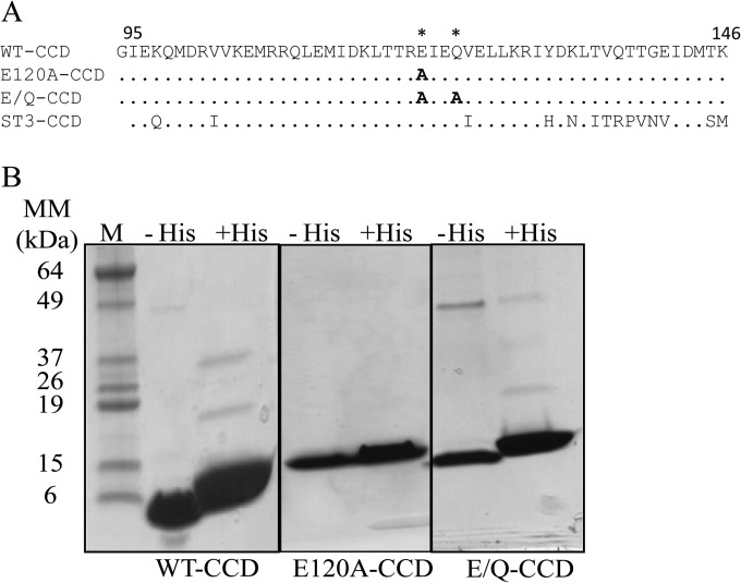
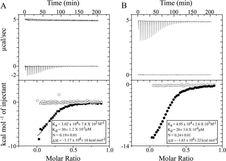
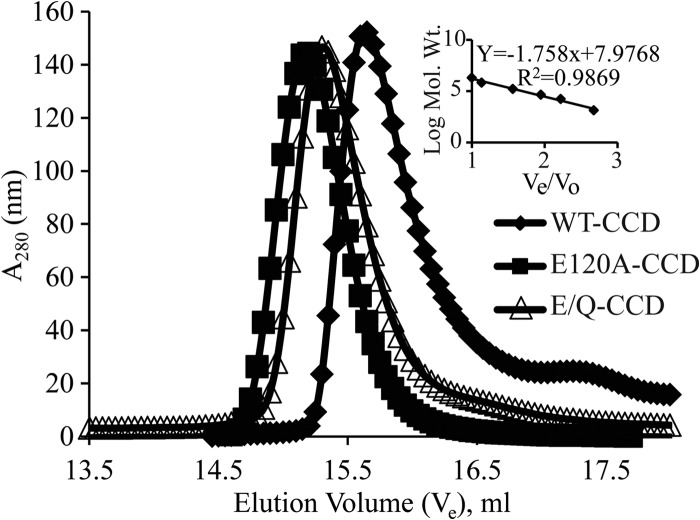
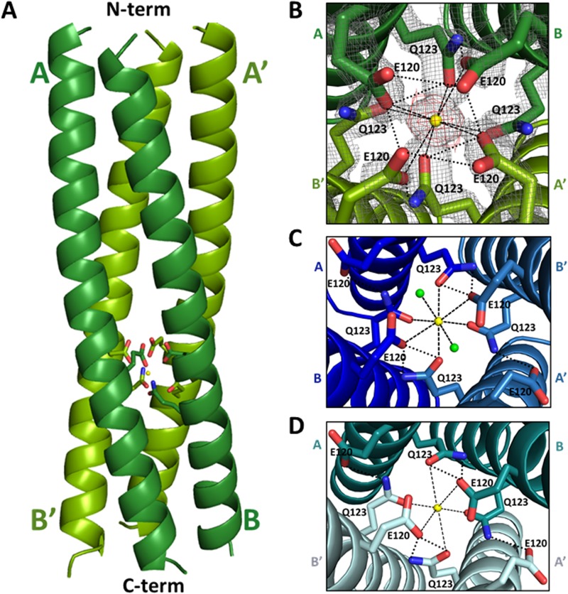
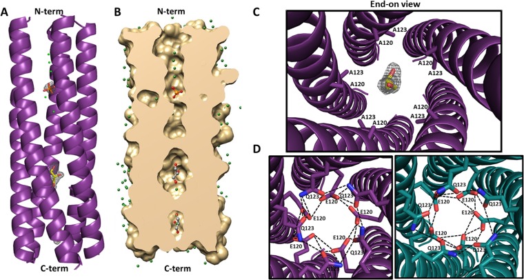
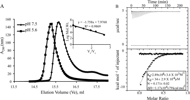
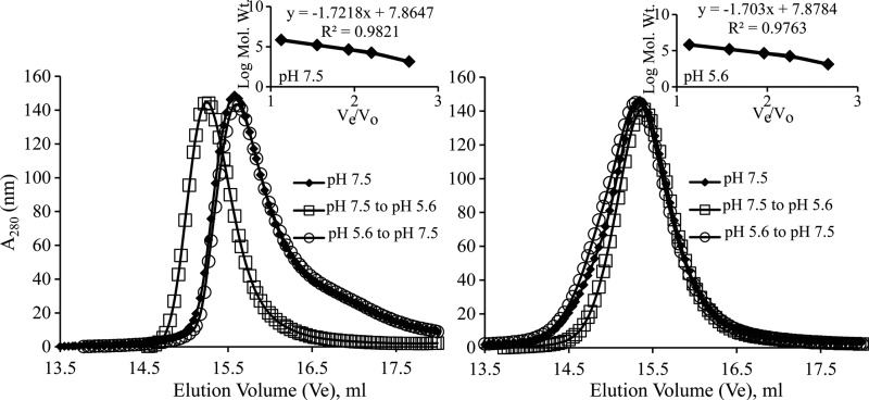
Rotavirus (RV) nonstructural protein 4 (NSP4) is a virulence factor that disrupts cellular Ca(2+) homeostasis and plays multiple roles regulating RV replication and the pathophysiology of RV-induced diarrhea. Although its native oligomeric state is unclear, crystallographic studies of the coiled-coil domain (CCD) of NSP4 from two different strains suggest that it functions as a tetramer or a pentamer. While the CCD of simian strain SA11 NSP4 forms a tetramer that binds Ca(2+) at its core, the CCD of human strain ST3 forms a pentamer lacking the bound Ca(2+) despite the residues (E120 and Q123) that coordinate Ca(2+) binding being conserved. In these previous studies, while the tetramer crystallized at neutral pH, the pentamer crystallized at low pH, suggesting that preference for a particular oligomeric state is pH dependent and that pH could influence Ca(2+) binding. Here, we sought to examine if the CCD of NSP4 from a single RV strain can exist in two oligomeric states regulated by Ca(2+) or pH. Biochemical, biophysical, and crystallographic studies show that while the CCD of SA11 NSP4 exhibits high-affinity binding to Ca(2+) at neutral pH and forms a tetramer, it does not bind Ca(2+) at low pH and forms a pentamer, and the transition from tetramer to pentamer is reversible with pH. Mutational analysis shows that Ca(2+) binding is necessary for the tetramer formation, as an E120A mutant forms a pentamer. We propose that the structural plasticity of NSP4 regulated by pH and Ca(2+) may form a basis for its pleiotropic functions during RV replication.

Importance: The nonstructural protein NSP4 of rotavirus is a multifunctional protein that plays an important role in virus replication, morphogenesis, and pathogenesis. Previous crystallography studies of the coiled-coil domain (CCD) of NSP4 from two different rotavirus strains showed two distinct oligomeric states, a Ca(2+)-bound tetrameric state and a Ca(2+)-free pentameric state. Whether NSP4 CCD from the same strain can exist in different oligomeric states and what factors might regulate its oligomeric preferences are not known. This study used a combination of biochemical, biophysical, and crystallography techniques and found that the NSP4 CCD can undergo a reversible transition from a Ca(2+)-bound tetramer to a Ca(2+)-free pentamer in response to changes in pH. From these studies, we hypothesize that this remarkable structural adaptability of the CCD forms a basis for the pleiotropic functional properties of NSP4.


Figures

FIG 1
FIG 1
Purification of NSP4 WT-CCD and the CCD mutants. (A) Sequence alignment of strain SA11 NSP4 residues 95 to 146 of WT-CCD and the E120A-CCD and E/Q-CCD mutants showing specific amino acid mutations in WT-CCD, indicated with asterisks. The strain ST3 NSP4 CCD is also shown in the alignment to show that the E120 and Q123 residues are conserved. (B) SDS-PAGE gel of the NSP4 WT-CCD, E120A-CCD, and E/Q-CCD purified proteins with His tag (+His) and without His tag (−His). Molecular mass marker bands (lane M) are shown on left of the gel.
FIG 2
FIG 2
ITC analysis of divalent metal ions binding to NSP4 WT-CCD and CCD mutants. (A) Top, raw data from titrations of a 0.2-ml cell containing 480 μM WT-CCD was titrated with 36 × 1 μl of 2 mM CaCl2 in 20 mM Tris and 150 mM NaCl at pH 7.5; bottom, ITC data showing integrated isotherm and best-associated fit for a one-site model showing Ca2+ binding to WT-CCD (■), E120A-CCD (○), and E/Q-CCD (△) proteins. Average thermodynamic parameters associated with Ca2+ binding to WT-CCD are reported in the inset. E120A-CCD and E/Q-CCD do not show any binding with Ca2+. (B) Top, raw data from titrations of a 0.2-ml cell containing 480 μM WT-CCD was titrated with 36 × 1 μl of 2 mM BaCl2 or MgCl2 in 20 mM Tris and 150 mM NaCl at pH 7.5; bottom, ITC data showing integrated isotherm and best-associated fit for a one-site model showing Ba2+ (●) and Mg2+ (□) binding to WT-CCD at pH 7.5. Average thermodynamic parameters associated with Ba2+ binding to WT-CCD at pH 7.5 are reported in the inset. At pH 7.5, no binding of Mg2+ is observed.
FIG 3
FIG 3
Effect of mutation of Ca2+ coordinating amino acids on oligomerization of the NSP4 CCD. Molecular mass determinations by size exclusion chromatography. A molecular mass calibration curve was obtained from the elution profiles of the standard proteins (inset). Apparent molecular masses of the proteins were determined from a standard graph. WT-CCD eluted as a tetramer (26.4 kDa) (◆), whereas mutants E120A-CCD (33.6 kDa) (■) and E/Q-CCD (31.1 kDa) (△) eluted as pentamers at pH 7.5. Elution volumes, apparent molecular masses, and numbers of oligomers are reported in Table 2.
FIG 4
FIG 4
Comparison of the Ca2+-binding sites in the SA11 NSP4 CCD structures. (A) A side view of the ribbon representation of the NSP4 CCD (residues 95 to 145) tetramer presented in this paper. The bound Ca2+ ion (yellow sphere) along with E120 and Q123 (rendered as sticks, with nitrogen shown in blue and oxygen shown in red) are shown. The tetramer is formed by the association of two parallel helices (A and B, shown in dark green) in the asymmetric unit in the crystal with their crystallographic 2-fold symmetry-related mates (A′ and B′; shown in light green). The helical nature of all four chains extends from residue 95 (labeled as N-term on the top) to 136 (labeled as C-term on the bottom), with the last 10 residues (137 to 145) not clearly resolved in the structure. (B) Closeup of the end-on view of the Ca2+-binding site, with E120 and Q123 residues shown as sticks and the Ca2+ ion shown as a yellow sphere. In this structure, Ca2+ ion binding is coordinated by four E120 and Q123 residues (rendered as sticks, with nitrogen shown in blue and oxygen shown in red). 2Fo-Fc density for the E120 and Q123 residues at a contour level of 1σ is shown in gray, and Fo-Fc difference map density for Ca2+ (3σ contour level) is shown in red. (C) Ca2+-binding site in the SA11 NSP4 CCD tetramer (PDB 1G1I) reported by Bowman et al. (13). A and B subunits in the asymmetric unit (dark blue) and their crystallographic 2-fold symmetry-related mates A′ and B′ (light blue) are shown. In this structure, Ca2+ ion (yellow sphere) binding is coordinated by two water molecules (shown as green spheres), two E120 residues, and four Q123 residues (rendered as sticks). (D) Ca2+-binding site in the SA11 NSP4 CCD tetramer (PDB 2O1K) reported by Deepa et al. (15). Two molecules in the asymmetric unit (A and B [dark teal]) and their crystallographic 2-fold symmetry-related mates (A′ and B′ [light teal]) are shown. Ca2+ ion (yellow sphere) binding is coordinated by two E120 residues and four Q123 residues (rendered as sticks). In panels B, C, and D, the ionic interactions with Ca2+ and hydrogen bond interactions between the side chains are shown as dashed and dotted black lines, respectively.
FIG 5
FIG 5
Structure of the E120A/Q123A SA11 NSP4 CCD pentamer. (A) A side view of the ribbon representation of E120A/Q123A SA11 NSP4 CCD (residues 95 to 146) pentamer formed by five parallel helices (purple). The helical nature of all five chains extends from residues 95 (labeled as N-term on the top) to 137 (labeled as C-term on the bottom), with the last nine residues not clearly resolved in the structure. A phosphate molecule (orange sticks with oxygen shown in red) and two glycerol molecules (yellow sticks with oxygen shown in red) are depicted inside the channel. Fo-Fc difference map densities (3σ level) for the phosphate and glycerol molecules are shown in gray. Water molecules are shown as green spheres. (B) A longitudinal slice of the E/Q-CCD pentamer surface model (tan) revealing a cross section of the axial pore. A phosphate molecule is rendered as orange sticks, and two glycerol molecules are rendered as gray sticks, with oxygen shown in red. Water molecules are shown as green spheres. (C) Closeup of the end-on view of the mutated Ca2+-binding site, with A120 and A123 residues shown as purple sticks, a glycerol molecule shown as yellow sticks, and oxygen shown in red. Difference map density for the glycerol molecule is shown in gray. (D) Modeling of the E120 and Q123 residues into the E/Q-CCD pentamer structure and comparison with the ST3 CCD pentamer (PDB 3MIW) reported by Chacko et al. (14). Right, alanine residues at positions 120 and 123 in the E/Q-CCD were mutated back to E and Q (purple sticks, with nitrogen shown in blue and oxygen shown in red), respectively, as in the native SA11 sequence. The diameter of the pentamer accommodates all 10 residues and allows for formation of hydrogen bond interactions (dotted black lines) between the residues that are very similar to the interactions formed in the ST3 pentamer. Left, closeup end-on view of the hydrogen bond interaction (dashed black lines) between E120 and Q123 residues in the ST3 CCD pentamer (dark teal). E120 and Q123 residues are rendered as teal sticks, with nitrogen and oxygen atoms shown in blue and red, respectively.
FIG 6
FIG 6
Effect of pH on Ca2+ binding and oligomerization of WT-CCD. (A) Molecular mass determinations by size exclusion chromatography. A molecular mass calibration curve was obtained from the elution profiles of the standard proteins (inset). Apparent molecular masses of the proteins were determined from a standard graph reported in Table 2. WT-CCD eluted as a 26.4-kDa oligomer at pH 7.5 (◆), consistent with a tetramer, whereas it eluted as 37.6 kDa at pH 5.6 (■), consistent with a larger oligomeric form (pentamer). (B) Top, raw data from titrations of a 0.2-ml cell containing 480 μM WT-CCD was titrated with 36 × 1 μl of 2 mM CaCl2 in 20 mM Tris and 150 mM NaCl at pH 7.5 and 20 mM NaAc and 150 mM NaCl at pH 5.6; bottom, ITC data showing integrated isotherm and best-associated fit for a one-site model showing Ca2+ binding to WT-CCD at pH 7.5 (stars) and at pH 5.6 (▼). Average thermodynamic parameters associated with Ca2+ binding to WT-CCD at pH 7.5 are reported in the inset. At pH 5.6, no binding of Ca2+ is observed.
FIG 7
FIG 7
Effect of pH on oligomer transition of NSP4 WT-CCD and E/Q-CCD. (A) Molecular mass determinations by size exclusion chromatography with varying pHs. WT-CCD eluted as a tetramer at pH 7.5 (◆ and ○), whereas it eluted as a larger oligomeric form as a pentamer at pH 5.6 (□). (B) E/Q-CCD eluted as a pentamer consistently at two different pH buffers (◆, □, and ○). A molecular mass calibration curve was obtained from the elution profiles of the standard proteins (inset). Apparent molecular masses of the proteins were determined from a standard graph (Table 2).

Similar articles

Cited by

References

    1. Zhou Y, Frey TK, Yang JJ. 2009. Viral calciomics: interplays between Ca2+ and virus. Cell Calcium 46:1–17. 10.1016/j.ceca.2009.05.005. - DOI - PMC - PubMed
    1. Bhowmick R, Halder UC, Chattopadhyay S, Chanda S, Nandi S, Bagchi P, Nayak MK, Chakrabarti O, Kobayashi N, Chawla-Sarkar M. 2012. Rotaviral enterotoxin nonstructural protein 4 targets mitochondria for activation of apoptosis during infection. J. Biol. Chem. 287:35004–35020. 10.1074/jbc.M112.369595. - DOI - PMC - PubMed
    1. Chattopadhyay S, Basak T, Nayak MK, Bhardwaj G, Mukherjee A, Bhowmick R, Sengupta S, Chakrabarti O, Chatterjee NS, Chawla-Sarkar M. 2013. Identification of cellular calcium binding protein calmodulin as a regulator of rotavirus A infection during comparative proteomic study. PLoS One 8:e56655. 10.1371/journal.pone.0056655. - DOI - PMC - PubMed
    1. Crawford SE, Hyser JM, Utama B, Estes MK. 2012. Autophagy hijacked through viroporin-activated calcium/calmodulin-dependent kinase kinase-beta signaling is required for rotavirus replication. Proc. Natl. Acad. Sci. U. S. A. 109:E3405–E3413. 10.1073/pnas.1216539109. - DOI - PMC - PubMed
    1. Diaz Y, Pena F, Aristimuno OC, Matteo L, De Agrela M, Chemello ME, Michelangeli F, Ruiz MC. 2012. Dissecting the Ca(2)(+) entry pathways induced by rotavirus infection and NSP4-EGFP expression in Cos-7 cells. Virus Res. 167:285–296. 10.1016/j.virusres.2012.05.012. - DOI - PubMed

Publication types

MeSH terms

LinkOut - more resources