. 2014 Dec;88(23):13759-68.
doi: 10.1128/JVI.02166-14. Epub 2014 Sep 17.

Molecular mimicry between dengue virus and coagulation factors induces antibodies to inhibit thrombin activity and enhance fibrinolysis

Affiliations

Molecular mimicry between dengue virus and coagulation factors induces antibodies to inhibit thrombin activity and enhance fibrinolysis

Yung-Chun Chuang et al. J Virol. 2014 Dec.

Abstract

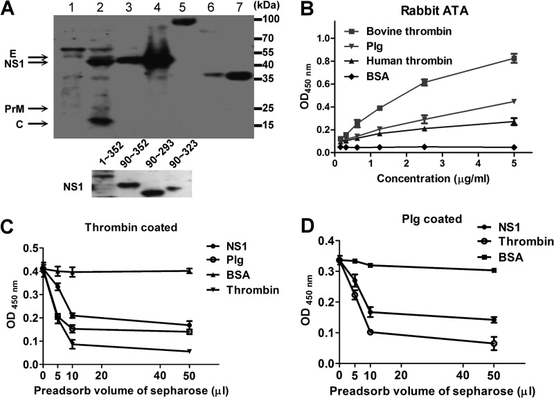
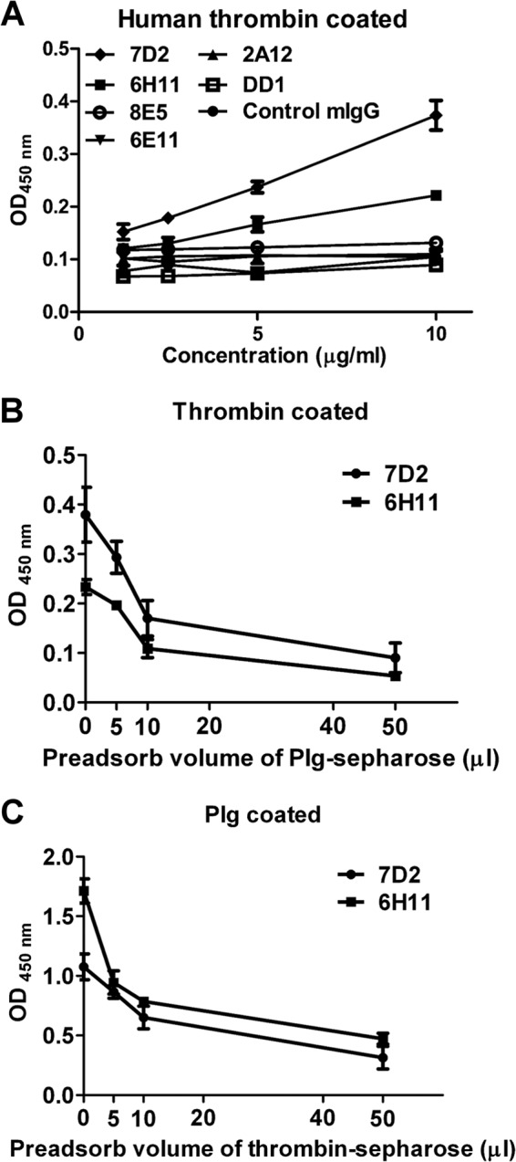
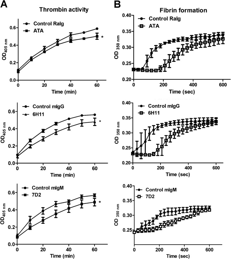
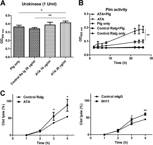
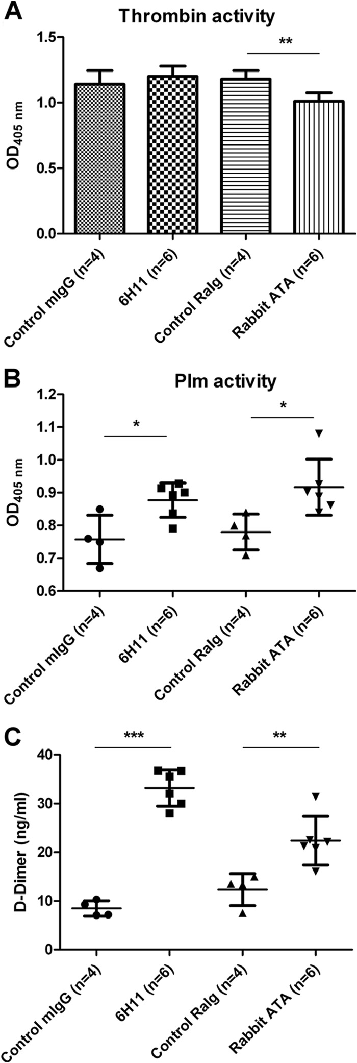
Dengue virus (DENV) is the most common cause of viral hemorrhagic fever, and it may lead to life-threating dengue hemorrhagic fever and shock syndrome (DHF/DSS). Because most cases of DHF/DSS occur in patients with secondary DENV infection, anti-DENV antibodies are generally considered to play a role in the pathogenesis of DHF/DSS. Previously, we have found that antithrombin antibodies (ATAs) with both antithrombotic and profibrinolytic activities are present in the sera of dengue patients. However, the mechanism by which these autoantibodies are induced is unclear. In this study, we demonstrated that antibodies induced by DENV immunization in mice and rabbits could bind to DENV antigens as well as to human thrombin and plasminogen (Plg). The binding of anti-DENV antibodies to thrombin and Plg was inhibited by preadsorption with DENV nonstructural protein 1. In addition, affinity-purified ATAs from DENV-immunized rabbit sera could inhibit thrombin activity and enhance Plg activation both in vitro and in vivo. Taken together, our results suggest that molecular mimicry between DENV and coagulation factors can induce the production of autoantibodies with biological effects similar to those of ATAs found in dengue patients. These coagulation-factor cross-reactive anti-DENV antibodies can interfere with the balance of coagulation and fibrinolysis, which may lead to the tendency of DHF/DSS patients to bleed.

Importance: Dengue virus (DENV) infection is the most common mosquito-borne viral disease in tropical and subtropical areas. Over 50 million DENV infection cases develop each year, and more than 2.5 billion people are at risk of dengue-induced hemorrhagic fever and shock syndrome. Currently, there is no vaccine or drug treatment for DENV. In the present study, we demonstrated that DENV immunization could induce thrombin and plasminogen (Plg) cross-reactive antibodies, which were able to inhibit thrombin activity and enhance Plg activation. These results suggest that molecular mimicry between DENV antigens, thrombin, and Plg may elicit antibodies that disturb hemostasis. The selection of appropriate candidate antigens for use in DENV vaccines should prevent these potentially dangerous autoimmune responses.


Figures

FIG 1
FIG 1
Antibodies against thrombin and other coagulation factors in DENV-immunized mouse sera. (A) Mice were immunized with DENV antigens as described in Materials and Methods. Serum samples were collected on day 32. Antibodies bound to DENV antigens, rNS1, and human thrombin were detected by ELISA. (B) Different concentrations of DENV antigens, rNS1, or BSA were preincubated with DENV-immunized mouse sera before their addition to human thrombin-coated ELISA plates. Bound antibodies were detected as described in Materials and Methods. (C) Antibodies in the DENV-immunized mouse sera were bound to various human coagulation factors, as determined using different coagulation factor-coated ELISA plates. The results are presented as the means ± standard deviations from three independent experiments.
FIG 2
FIG 2
Characterization of rabbit ATAs. (A) Western blotting was performed to assess the ability of the rabbit ATAs to bind to DENV protein, Plg, human/bovine thrombin (top panel), and recombinant NS1 proteins of different lengths as indicated (bottom panel). Lane 1, C6/36 cell lysate; lane 2, DENV-infected C6/36 cell lysate; lane 3, supernatants from DENV-infected C6/36 cells; lane 4, PEG-precipitated DENV antigens; lane 5, Plg; lane 6, bovine thrombin; lane 7, human thrombin. (B) Ability of rabbit ATAs to bind to bovine/human thrombin, Plg, and BSA as determined by ELISA. (C and D) Rabbit ATAs were preadsorbed to BSA-, NS1-, Plg-, or thrombin-conjugated Sepharose as indicated. The ability of the nonadsorbed antibodies to bind to thrombin or Plg was determined by ELISA. The results are presented as the means ± standard deviations from three independent experiments.
FIG 3
FIG 3
Anti-DENV MAbs cross-react with both Plg and thrombin. (A) The ability of the Plg cross-reactive MAbs to bind to human thrombin was determined by ELISA. (B) MAbs 7D2 and 6H11 (10 μg/ml) were used for preadsorption to Plg-conjugated Sepharose. (C) Five micrograms per milliliter of each MAb was used for thrombin-conjugated Sepharose preadsorption. The binding of MAbs 7D2 and 6H11 to human thrombin or Plg was detected by ELISA. The results are presented as the means ± standard deviations from three independent experiments.
FIG 4
FIG 4
ATAs and MAbs from DENV-immunized animals inhibit thrombin activity in vitro. (A) Human thrombin was preincubated with ATAs, MAbs (7D2 and 6H11), control rabbit Igs (RaIg), control mouse IgG (mIgG), or control mouse IgM (mIgM) for 1 h, and thrombin activity was determined by S-2238. (B) Human thrombin was preincubated with antibodies for 1 h before its addition to human PPP. Fibrin formation was detected by the change in turbidity at OD350. The data are presented as the means ± standard deviations from three independent experiments. Significance was analyzed by two-way ANOVA (*, P < 0.05).
FIG 5
FIG 5
ATAs and MAbs from DENV-immunized animals enhance Plg activation and fibrinolysis in vitro. (A) Plg was incubated with control rabbit Igs or ATAs before its addition to urokinase and S-2251. After 1 h of incubation, Plm formation was measured by monitoring the OD at 405 nm. (B) S-2251 was coincubated with Plg, ATAs, control rabbit Igs, or Plg with ATAs for different time periods as indicated. Plm formation was determined as described above. (C) Human PPP was incubated with control rabbit Igs or with ATAs and urokinase before the addition of human thrombin. The percentage of clot lysis was calculated as described in Materials and Methods. The results are presented as the means ± standard deviations from three independent experiments (*, P < 0.5; **, P < 0.01; Student's t test).
FIG 6
FIG 6
Effects of ATAs and MAb 6H11 on thrombin activity and fibrinolysis in vivo. (A) Control mouse IgG (n = 4), control rabbit Igs (n = 4), MAb 6H11 (n = 6), or ATAs (n = 6) were injected intravenously into ICR mice (5 μg/g body weight; average weight of 20 to 25 g). Thrombin activity in the mouse PPP was determined by S-2238 as described in Materials and Methods. (B) Plm activity of the mouse PPP was measured by S-2251 as described in Materials and Methods. (C) The levels of d-dimer in the mouse PPP were measured by competition ELISA as described in Materials and Methods. The results are presented as the means ± standard deviations from three independent experiments (*, P < 0.5; **, P < 0.01; ***, P < 0.005; Student's t test).
FIG 7
FIG 7
Epitope analysis of MAbs 6H11 and 7D2. (A) Consensus sequence analysis comparing MAb 6H11 and 7D2 epitopes with DENV proteins, human thrombin, and Plg. White text on black background indicates complete homology. Black text on gray background depicts phage and antigen amino acids with similar characteristics. (B) MAb 6H11 or 7D2 was preincubated with its epitope phage (cluster 1 for MAb 6H11 and cluster 3 for MAb 7D2) or helper phage for 1 h. Their ability to bind to human thrombin was determined by ELISA. The data are presented as the means ± standard deviations from three independent experiments. Significance was analyzed by two-way ANOVA (**, P < 0.01).

Similar articles

Cited by

References

    1. Guzman MG, Halstead SB, Artsob H, Buchy P, Farrar J, Gubler DJ, Hunsperger E, Kroeger A, Margolis HS, Martinez E, Nathan MB, Pelegrino JL, Simmons C, Yoksan S, Peeling RW. 2010. Dengue: a continuing global threat. Nat. Rev. Microbiol. 8:S7–S16. 10.1038/nrmicro2460. - DOI - PMC - PubMed
    1. Young PR, Hilditch PA, Bletchly C, Halloran W. 2000. An antigen capture enzyme-linked immunosorbent assay reveals high levels of the dengue virus protein NS1 in the sera of infected patients. J. Clin. Microbiol. 38:1053–1057. - PMC - PubMed
    1. Alcon S, Talarmin A, Debruyne M, Falconar A, Deubel V, Flamand M. 2002. Enzyme-linked immunosorbent assay specific to dengue virus type 1 nonstructural protein NS1 reveals circulation of the antigen in the blood during the acute phase of disease in patients experiencing primary or secondary infections. J. Clin. Microbiol. 40:376–381. 10.1128/JCM.40.02.376-381.2002. - DOI - PMC - PubMed
    1. Guzman MG, Kouri G. 2002. Dengue: an update. Lancet Infect. Dis. 2:33–42. 10.1016/S1473-3099(01)00171-2. - DOI - PubMed
    1. Halstead SB. 1982. Dengue: hematologic aspects. Semin. Hematol. 19:116–131. - PubMed

Publication types