

Molecular characterization of common fragile sites as a strategy to discover cancer susceptibility genes
- PMID: 25231336
- PMCID: PMC11114050
- DOI: 10.1007/s00018-014-1723-z
Molecular characterization of common fragile sites as a strategy to discover cancer susceptibility genes
Abstract
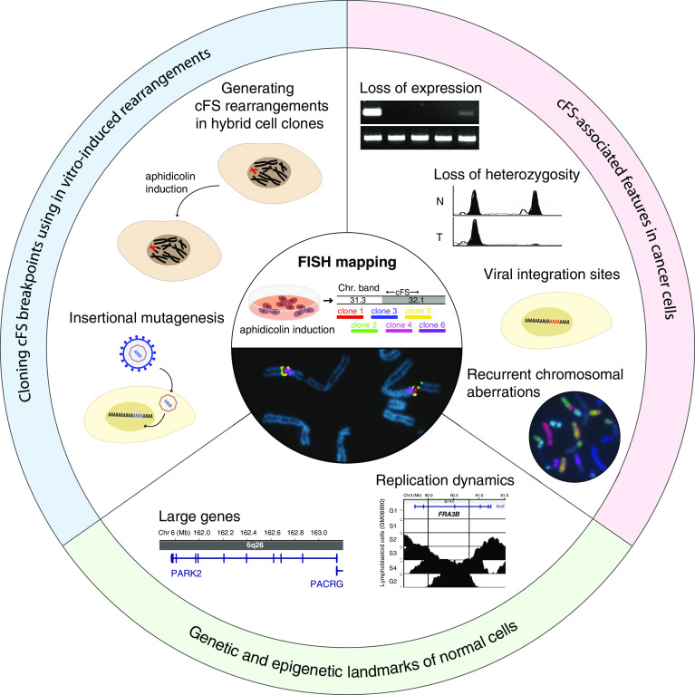
The cytogenetic hypothesis that common fragile sites (cFSs) are hotspots of cancer breakpoints is increasingly supported by recent data from whole-genome profiles of different cancers. cFSs are components of the normal chromosome structure that are particularly prone to breakage under conditions of replication stress. In recent years, cFSs have become of increasing interest in cancer research, as they not only appear to be frequent targets of genomic alterations in progressive tumors, but also already in precancerous lesions. Despite growing evidence of their importance in disease development, most cFSs have not been investigated at the molecular level and most cFS genes have not been identified. In this review, we summarize the current data on molecularly characterized cFSs, their genetic and epigenetic characteristics, and put emphasis on less-studied cFS genes as potential contributors to cancer development.
Conflict of interest statement
The authors declare that they have no conflicts of interest.
Figures

Similar articles
-
Impaired Replication Timing Promotes Tissue-Specific Expression of Common Fragile Sites.Genes (Basel). 2020 Mar 19;11(3):326. doi: 10.3390/genes11030326. Genes (Basel). 2020. PMID: 32204553 Free PMC article.
-
Interplay between genetic and epigenetic factors governs common fragile site instability in cancer.Cell Mol Life Sci. 2014 Dec;71(23):4495-506. doi: 10.1007/s00018-014-1719-8. Epub 2014 Oct 9. Cell Mol Life Sci. 2014. PMID: 25297918 Free PMC article. Review.
-
Common fragile sites are conserved features of human and mouse chromosomes and relate to large active genes.Genome Res. 2006 Oct;16(10):1222-30. doi: 10.1101/gr.5335506. Epub 2006 Sep 5. Genome Res. 2006. PMID: 16954539 Free PMC article.
-
Large transcription units unify copy number variants and common fragile sites arising under replication stress.Genome Res. 2015 Feb;25(2):189-200. doi: 10.1101/gr.177121.114. Epub 2014 Nov 4. Genome Res. 2015. PMID: 25373142 Free PMC article.
-
Updating the mechanisms of common fragile site instability: how to reconcile the different views?Cell Mol Life Sci. 2014 Dec;71(23):4489-94. doi: 10.1007/s00018-014-1720-2. Epub 2014 Sep 24. Cell Mol Life Sci. 2014. PMID: 25248392 Free PMC article. Review.
Cited by
-
WWOX modulates the ATR-mediated DNA damage checkpoint response.Oncotarget. 2016 Jan 26;7(4):4344-55. doi: 10.18632/oncotarget.6571. Oncotarget. 2016. PMID: 26675548 Free PMC article.
-
Replication Stress Induces Global Chromosome Breakage in the Fragile X Genome.Cell Rep. 2020 Sep 22;32(12):108179. doi: 10.1016/j.celrep.2020.108179. Cell Rep. 2020. PMID: 32966779 Free PMC article.
-
Epigenomic signatures associated with spontaneous and replication stress-induced DNA double strand breaks.Front Genet. 2022 Nov 24;13:907547. doi: 10.3389/fgene.2022.907547. eCollection 2022. Front Genet. 2022. PMID: 36506300 Free PMC article.
-
DNA secondary structure at chromosomal fragile sites in human disease.Curr Genomics. 2015 Feb;16(1):60-70. doi: 10.2174/1389202916666150114223205. Curr Genomics. 2015. PMID: 25937814 Free PMC article.
-
Fragile sites, chromosomal lesions, tandem repeats, and disease.Front Genet. 2022 Nov 17;13:985975. doi: 10.3389/fgene.2022.985975. eCollection 2022. Front Genet. 2022. PMID: 36468036 Free PMC article. Review.
References
-
- Lukusa T, Fryns JP. Human chromosome fragility. Biochim Biophys Acta. 2008;1779(1):3–16. - PubMed
-
- Gorgoulis VG, Vassiliou LV, Karakaidos P, Zacharatos P, Kotsinas A, Liloglou T, Venere M, Ditullio RA, Jr, Kastrinakis NG, Levy B, Kletsas D, Yoneta A, Herlyn M, Kittas C, Halazonetis TD. Activation of the DNA damage checkpoint and genomic instability in human precancerous lesions. Nature. 2005;434(7035):907–913. - PubMed
-
- Halazonetis TD, Gorgoulis VG, Bartek J. An oncogene-induced DNA damage model for cancer development. Science. 2008;319(5868):1352–1355. - PubMed
-
- Durkin SG, Glover TW. Chromosome fragile sites. Annu Rev Genet. 2007;41:169–192. - PubMed
-
- Berger R, Bloomfield CD, Sutherland GR. Report of the committee on chromosome rearrangements in neoplasia and on fragile sites. Cytogenet Cell Genet. 1985;40(1–4):490–535. - PubMed
Publication types
MeSH terms
Substances
LinkOut - more resources
Full Text Sources
Other Literature Sources
