. 2015 Feb;21(3-4):637-46.
doi: 10.1089/ten.TEA.2013.0679. Epub 2014 Oct 23.

Delivering heparin-binding insulin-like growth factor 1 with self-assembling peptide hydrogels

Affiliations

Delivering heparin-binding insulin-like growth factor 1 with self-assembling peptide hydrogels

Emily M Florine et al. Tissue Eng Part A. 2015 Feb.

Abstract

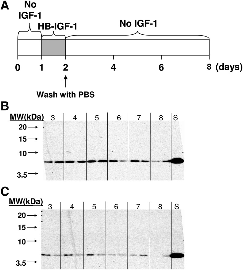
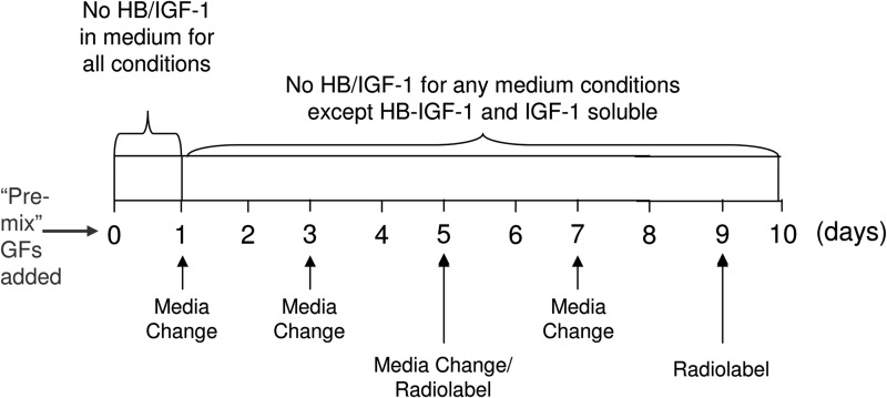
Heparin-binding insulin-like growth factor 1 (HB-IGF-1) is a fusion protein of IGF-1 with the HB domain of heparin-binding epidermal growth factor-like growth factor. A single dose of HB-IGF-1 has been shown to bind specifically to cartilage and to promote sustained upregulation of proteoglycan synthesis in cartilage explants. Achieving strong integration between native cartilage and tissue-engineered cartilage remains challenging. We hypothesize that if a growth factor delivered by the tissue engineering scaffold could stimulate enhanced matrix synthesis by both the cells within the scaffold and the adjacent native cartilage, integration could be enhanced. In this work, we investigated methods for adsorbing HB-IGF-1 to self-assembling peptide hydrogels to deliver the growth factor to encapsulated chondrocytes and cartilage explants cultured with growth factor-loaded hydrogels. We tested multiple methods for adsorbing HB-IGF-1 in self-assembling peptide hydrogels, including adsorption prior to peptide assembly, following peptide assembly, and with/without heparan sulfate (HS, a potential linker between peptide molecules and HB-IGF-1). We found that HB-IGF-1 and HS were retained in the peptide for all tested conditions. A subset of these conditions was then studied for their ability to stimulate increased matrix production by gel-encapsulated chondrocytes and by chondrocytes within adjacent native cartilage. Adsorbing HB-IGF-1 or IGF-1 prior to peptide assembly was found to stimulate increased sulfated glycosaminoglycan per DNA and hydroxyproline content of chondrocyte-seeded hydrogels compared with basal controls at day 10. Cartilage explants cultured adjacent to functionalized hydrogels had increased proteoglycan synthesis at day 10 when HB-IGF-1 was adsorbed, but not IGF-1. We conclude that delivery of HB-IGF-1 to focal defects in cartilage using self-assembling peptide hydrogels is a promising technique that could aid cartilage repair via enhanced matrix production and integration with native tissue.


Figures

<b>FIG. 1.</b>
FIG. 1.
Heparin-binding insulin-like growth factor 1 (HB-IGF-1) dose–response in cartilage explants at day 8 following wash-out procedure. (A) Timeline of experimental procedure. Explants were treated with 50 nM IGF-1, or 0.5, 5, 50, or 100 nM HB-IGF-1 from day 0 to 2. (B) Proteoglycan synthesis normalized to DNA content (μg) and the no-growth factor (“0”) condition (dashed line) at day 8. Values are mean±standard error of the mean (SEM). N=6 cartilage disks per condition, one animal. Solid lines indicate significant difference between two conditions, p<0.05. Color images available online at www.liebertpub.com/tea
<b>FIG. 2.</b>
FIG. 2.
Heparan sulfate (HS) release from acellular peptide hydrogels. Peptide with 5 μM HS cultured in 100 μL bath of phosphate-buffered saline (PBS), PBS with 1 M NaCl (“1 M NaCl), or PBS with a pH of 9 (adjusted using NaOH, “pH 9”). Values are mean±SEM. N=6–8, except at day 6 where n=2. Versus PBS alone, *versus pH 9, p<0.05. Color images available online at www.liebertpub.com/tea
<b>FIG. 3.</b>
FIG. 3.
Growth factor retention when mixed into initially unassembled acellular peptide. Western blots of (A) HB-IGF-1 and (B) IGF-1 retained in peptide with or without HS on days 1 through 6, using an anti-IGF-1 antibody. Std is a standard of 5 ng HB-IGF-1 or IGF-1 for (A) and (B), respectively.
<b>FIG. 4.</b>
FIG. 4.
HB-IGF-1 retention when adsorbed to preassembled acellular peptide. (A) Time course of experimental procedure where peptide±HS assembled from day 0 to 1 and HB-IGF-1 was absorbed to peptide from day 1 to 2. Western blots of (B) HB-IGF-1 retained by days 3–8 after adsorption to peptide alone, and (C) HB-IGF-1 retained by days 3–8 after adsorption to peptide mixed with HS prior to assembly. S is a standard of 5 ng HB-IGF-1. Two lanes for each time point are repeats of the same condition.
<b>FIG. 5.</b>
FIG. 5.
14C-IGF-1 and 14C-HB-IGF-1 release from acellular peptide scaffold. Peptide with 615 nM labeled growth factor was cultured in a 500 μL bath of PBS. Values are mean±standard deviation. N=4. Color images available online at www.liebertpub.com/tea
<b>FIG. 6.</b>
FIG. 6.
Time course of experimental procedures for bovine chondrocytes in regular or functionalized peptide hydrogels. Identical time course was used for cartilage explants cultured with functionalized peptide gels, where explants were harvested at day-1, received a media change on day 1, and were added to gel cultures on day 2.
<b>FIG. 7.</b>
FIG. 7.
Extracellular matrix content and biosynthesis by bovine chondrocytes encapsulated within peptide hydrogels. (A) Sulfated glycosaminoglycan (sGAG) content normalized by DNA content, (B) proteoglycan synthesis, (C) protein synthesis, (D) hydroxyproline content, and (E) key of culture conditions. Values are mean±SEM. N=5–6×2 experiments with cells from different animal donors; data from one experiment shown above. Line indicates significant difference between two conditions consistent between both experiments, p<0.05. See Figure 6 for time course of experimental procedure. Color images available online at www.liebertpub.com/tea
<b>FIG. 8.</b>
FIG. 8.
Proteoglycan synthesis within cartilage explants cultured with growth factor-functionalized acellular peptide hydrogel. Data normalized to DNA content. Values are mean±SEM. N=11–12 (3–4 explants×3 animals). Line indicates significant difference between two conditions, p<0.05. Color images available online at www.liebertpub.com/tea

References

    1. Ahmed T.A., and Hincke M.T.Strategies for articular cartilage lesion repair and functional restoration. Tissue Eng Part B Rev 16,305, 2010 - PubMed
    1. Anderson D.D., Chubinskaya S., Guilak F., Martin J.A., Oegema T.R., Olson S.A., and Buckwalter J.A.Post-traumatic osteoarthritis: improved understanding and opportunities for early intervention. J Orthop Res 29,802, 2011 - PMC - PubMed
    1. Tyler J.A.Insulin-like growth factor 1 can decrease degradation and promote synthesis of proteoglycan in cartilage exposed to cytokines. Biochem J 260,543, 1989 - PMC - PubMed
    1. Luyten F.P., Hascall V.C., Nissley S.P., Morales T.I., and Reddi A.H.Insulin-like growth factors maintain steady-state metabolism of proteoglycans in bovine articular cartilage explants. Arch Biochem Biophys 267,416, 1988 - PubMed
    1. McQuillan D.J., Handley C.J., Campbell M.A., Bolis S., Milway V.E., and Herington A.C.Stimulation of proteoglycan biosynthesis by serum and insulin-like growth factor-I in cultured bovine articular cartilage. Biochem J 240,423, 1986 - PMC - PubMed

Publication types

MeSH terms