Suppression of mTORC1 activation in acid-α-glucosidase-deficient cells and mice is ameliorated by leucine supplementation
- PMID: 25231351
- PMCID: PMC4233288
- DOI: 10.1152/ajpregu.00212.2014
Suppression of mTORC1 activation in acid-α-glucosidase-deficient cells and mice is ameliorated by leucine supplementation
Abstract
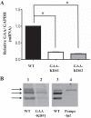
Pompe disease is due to a deficiency in acid-α-glucosidase (GAA) and results in debilitating skeletal muscle wasting, characterized by the accumulation of glycogen and autophagic vesicles. Given the role of lysosomes as a platform for mTORC1 activation, we examined mTORC1 activity in models of Pompe disease. GAA-knockdown C2C12 myoblasts and GAA-deficient human skin fibroblasts of infantile Pompe patients were found to have decreased mTORC1 activation. Treatment with the cell-permeable leucine analog L-leucyl-L-leucine methyl ester restored mTORC1 activation. In vivo, Pompe mice also displayed reduced basal and leucine-stimulated mTORC1 activation in skeletal muscle, whereas treatment with a combination of insulin and leucine normalized mTORC1 activation. Chronic leucine feeding restored basal and leucine-stimulated mTORC1 activation, while partially protecting Pompe mice from developing kyphosis and the decline in muscle mass. Leucine-treated Pompe mice showed increased spontaneous activity and running capacity, with reduced muscle protein breakdown and glycogen accumulation. Together, these data demonstrate that GAA deficiency results in reduced mTORC1 activation that is partly responsible for the skeletal muscle wasting phenotype. Moreover, mTORC1 stimulation by dietary leucine supplementation prevented some of the detrimental skeletal muscle dysfunction that occurs in the Pompe disease mouse model.
Keywords: Pompe disease; leucine; lysosome; mTORC1; muscle; α-glucosidase.
Copyright © 2014 the American Physiological Society.
Figures


Similar articles
-
Akt inactivation induces endoplasmic reticulum stress-independent autophagy in fibroblasts from patients with Pompe disease.Mol Genet Metab. 2012 Nov;107(3):490-5. doi: 10.1016/j.ymgme.2012.09.011. Epub 2012 Sep 15. Mol Genet Metab. 2012. PMID: 23041259
-
Glycosylation-independent lysosomal targeting of acid α-glucosidase enhances muscle glycogen clearance in pompe mice.J Biol Chem. 2013 Jan 18;288(3):1428-38. doi: 10.1074/jbc.M112.438663. Epub 2012 Nov 27. J Biol Chem. 2013. PMID: 23188827 Free PMC article.
-
Remarkably low fibroblast acid α-glucosidase activity in three adults with Pompe disease.Mol Genet Metab. 2012 Nov;107(3):485-9. doi: 10.1016/j.ymgme.2012.09.003. Epub 2012 Sep 7. Mol Genet Metab. 2012. PMID: 23000108
-
Autophagy in skeletal muscle: implications for Pompe disease.Int J Clin Pharmacol Ther. 2009;47 Suppl 1(Suppl 1):S42-7. doi: 10.5414/cpp47042. Int J Clin Pharmacol Ther. 2009. PMID: 20040311 Free PMC article. Review.
-
Gene Therapy for Pompe Disease: The Time is now.Hum Gene Ther. 2019 Oct;30(10):1245-1262. doi: 10.1089/hum.2019.109. Epub 2019 Sep 9. Hum Gene Ther. 2019. PMID: 31298581 Review.
Cited by
-
RAP1A suppresses hepatic steatosis by regulating amino acid-mediated mTORC1 activation.JHEP Rep. 2024 Dec 18;7(4):101303. doi: 10.1016/j.jhepr.2024.101303. eCollection 2025 Apr. JHEP Rep. 2024. PMID: 40124164 Free PMC article.
-
Modulation of mTOR signaling as a strategy for the treatment of Pompe disease.EMBO Mol Med. 2017 Mar;9(3):353-370. doi: 10.15252/emmm.201606547. EMBO Mol Med. 2017. PMID: 28130275 Free PMC article.
-
From Acid Alpha-Glucosidase Deficiency to Autophagy: Understanding the Bases of POMPE Disease.Int J Mol Sci. 2023 Aug 5;24(15):12481. doi: 10.3390/ijms241512481. Int J Mol Sci. 2023. PMID: 37569856 Free PMC article.
-
Pompe disease: Shared and unshared features of lysosomal storage disorders.Rare Dis. 2015 Jul 15;3(1):e1068978. doi: 10.1080/21675511.2015.1068978. eCollection 2015. Rare Dis. 2015. PMID: 26619007 Free PMC article.
-
Lysosomal glucose sensing and glycophagy in metabolism.Trends Endocrinol Metab. 2023 Nov;34(11):764-777. doi: 10.1016/j.tem.2023.07.008. Epub 2023 Aug 24. Trends Endocrinol Metab. 2023. PMID: 37633800 Free PMC article. Review.
References
-
- Agrawal S, Thakur P, Katoch SS. Beta adrenoceptor agonists, clenbuterol, and isoproterenol retard denervation atrophy in rat gastrocnemius muscle: use of 3-methylhistidine as a marker of myofibrillar degeneration. Jpn J Physiol 53: 229–237, 2003. - PubMed
-
- Ausems M, Verbiest J, Hermans M, Kroos M, Beemer F, Wokke J, Sandkuijl L, Reuser A, Van der Ploeg A. Frequency of glycogen storage disease type II in The Netherlands: implications for diagnosis and genetic counselling. Eur J Hum Genet 7: 713, 1999. - PubMed
-
- Bentzinger CF, Romanino K, Cloetta D, Lin S, Mascarenhas JB, Oliveri F, Xia J, Casanova E, Costa CF, Brink M, Zorzato F, Hall MN, Ruegg MA. Skeletal muscle-specific ablation of raptor, but not of rictor, causes metabolic changes and results in muscle dystrophy. Cell Metab 8: 411–424, 2008. - PubMed
-
- Case LE, Kishnani PS. Physical therapy management of Pompe disease. Genet Med 8: 318–327, 2006. - PubMed
-
- Castro AF, Rebhun JF, Clark GJ, Quilliam LA. Rheb binds tuberous sclerosis complex 2 (TSC2) and promotes S6 kinase activation in a rapamycin-and farnesylation-dependent manner. J Biol Chem 278: 32493–32496, 2003. - PubMed
Publication types
MeSH terms
Substances
Grants and funding
LinkOut - more resources
Full Text Sources
Other Literature Sources
Medical
Molecular Biology Databases
Miscellaneous

