Review
. 2014 Sep 17:9:37.
doi: 10.1186/1750-1326-9-37.

Chronic traumatic encephalopathy: clinical-biomarker correlations and current concepts in pathogenesis

Affiliations
Review

Chronic traumatic encephalopathy: clinical-biomarker correlations and current concepts in pathogenesis

Sam Gandy et al. Mol Neurodegener. .

Abstract

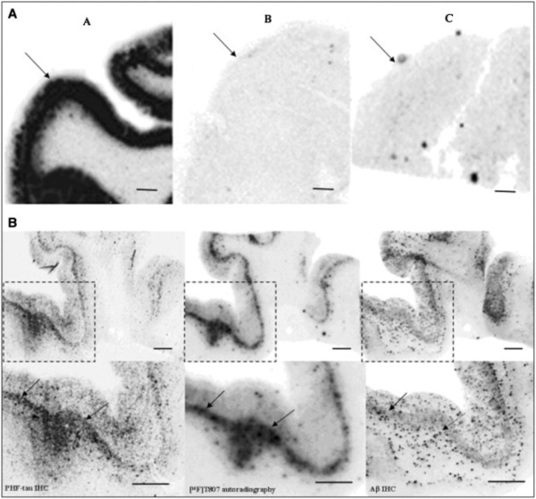
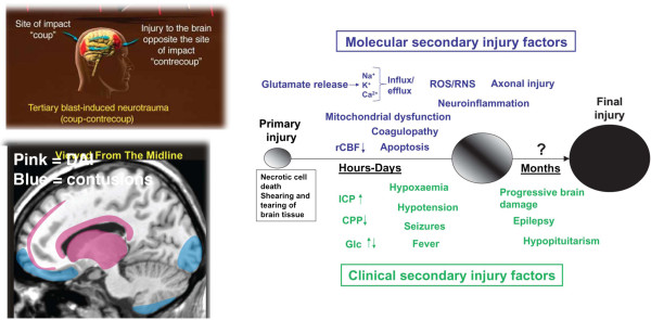
Background: Chronic traumatic encephalopathy (CTE) is a recently revived term used to describe a neurodegenerative process that occurs as a long term complication of repetitive mild traumatic brain injury (TBI). Corsellis provided one of the classic descriptions of CTE in boxers under the name "dementia pugilistica" (DP). Much recent attention has been drawn to the apparent association of CTE with contact sports (football, soccer, hockey) and with frequent battlefield exposure to blast waves generated by improvised explosive devices (IEDs). Recently, a promising serum biomarker has been identified by measurement of serum levels of the neuronal microtubule associated protein tau. New positron emission tomography (PET) ligands (e.g., [18 F] T807) that identify brain tauopathy have been successfully deployed for the in vitro and in vivo detection of presumptive tauopathy in the brains of subjects with clinically probable CTE.

Methods: Major academic and lay publications on DP/CTE were reviewed beginning with the 1928 paper describing the initial use of the term CTE by Martland.

Results: The major current concepts in the neurological, psychiatric, neuropsychological, neuroimaging, and body fluid biomarker science of DP/CTE have been summarized. Newer achievements, such as serum tau and [18 F] T807 tauopathy imaging, are also introduced and their significance has been explained.

Conclusion: Recent advances in the science of DP/CTE hold promise for elucidating a long sought accurate determination of the true prevalence of CTE. This information holds potentially important public health implications for estimating the risk of contact sports in inflicting permanent and/or progressive brain damage on children, adolescents, and adults.


Figures

Figure 1
Figure 1
Patterns of tau immunostaining in the frontal cortex of the patient with dementia pugilistica (DP), compared to Alzheimer’s disease (AD) and nondemented cases. MC-1 immunolabeling of phosphorylated tau revealed neuronal labeling in the DP case (A), plaque-associated neuritic labeling in the frontotemporal dementia (FTD)-AD case (B), a mix of neurofibrillary tangles (NFTs) and dystrophic neurites in the typical AD case (C), but absent in the control case (D). AT8 immunoreactivity also was limited to intracellular NFTs in the DP case (E), and within NFTs and plaque-associated dystrophic neurites in the FTD-AD case (F) and the typical AD case (G), but were not present in the control case (H). PHF-1 immunostaining was the most extensive of the three pathological tau markers, and showed significant intracellular and extracellular NFT labeling in the DP case (I), and within NFTs and plaque‐associated dystrophic neurites in the FTD-AD case (J), but was less extensive and limited to NFTs in the typical AD case (K). No PHF‐1-positive tangles were observed in the control case (L; scale bar = 100 μm). From Saing et al.[5] with permission.
Figure 2
Figure 2
Frontal cortical beta-amyloid (Aβ) neuropathology in dementia pugilistica (DP) as compared to that of Alzheimer’s disease (AD) and nondemented control cases. Aβ1-16 immunostaining illustrates primarily diffuse plaque and extracellular neurofibrillary tangle (NFT) labeling in the DP case (A) as compared to the extensive plaque labeling seen in the frontotemporal dementia (FTD)-­AD case (B), and the typical AD case (C), but is absent in the control case (D). Aβ1-42 immunostaining in the DP case (E), the FTD-AD case (F), the typical AD case (G), and the control (H), was similar to that observed with immunolabeling for Aβ1-16. Less Aβ1-40 immunolabeling was observed in the DP case (I), with deposits being primarily seen within diffuse plaques and on extracellular NFTs. In comparison, Aβ1-40 was observed in plaques in the FTD-AD case (J), and primarily within neuritic plaque cores and associated with blood vessels in the typical AD case (K), and was absent in the control case (L; scale bar = 500 μm). From Saing et al.[5] with permission.
Figure 3
Figure 3
Micrographs from the index case of CTE in an American football player. Panel A, β‐amyloid immunostain of the neocortex (original magnification, ×200) showing frequent diffuse amyloid plaques. Panel B, tau immunostain of the neocortex (original magnification, ×200) showing sparse NFTs and many tau-positive neuritic threads. Panel C, tau immunostain (original magnification, ×400) showing an NFT in a neocortical neuron with extending tau-positive dendritic processes. Panel D, β-amyloid immunostain (original magnification, ×100) of the Sommer’s sector (CA-1 region of the hippocampus) showing no diffuse amyloid plaques. From Omalu et al.[10] with permission.
Figure 4
Figure 4
The index case of military CTE. Photomicrographs of tau-immunostained section of the frontal cortex showing frequent neurofibrillary tangles and neuritic threads (A and B), with higher magnification (C and D) showing band- and flame-shaped neurofibrillary tangles and neuropil neuritic threads. Original magnification × 200 (A), × 400 (B), × 600 (C and D). From Omalu et al. [15] with permission.
Figure 5
Figure 5
[18 F] T807 autoradiography on brain sections and its comparison with paired helical filament (PHF)-tau and amyloid beta (Aβ) double immunohistochemistry (IHC). (A) Representative images for [18 F] T807 autoradiographs from groups A, B, and C of brains ([18 F] T807 autoradiography, 20 μCi/section). Positive autoradiography signals were observed only in the gray matter of brain from the PHF tau rich group A. Arrows indicate gray matter. (B) [18 F] T807 colocalized with PHF-tau but not with Aβ plaques. (B, top row) Low magnification. (B, bottom row) High magnification from the framed areas. Images of PHF tau (left) and Aβ (right) IHC double immunostaining and autoradiogram image (middle) from two adjacent sections (10 μm) from a PHF-tau rich group A brain (frontal lobe). Positive [18 F] T807 labeling colocalized with immunostaining of PHF tau but not with Aβ plaques, as indicated by arrows. Fluorescent and autoradiographic images were obtained using a Fuji Film FLA-7000 imaging instrument. Scale bars = 2 mm. From Xia et al.[19] with permission.
Figure 6
Figure 6
Molecular pathogenesis of TBI and CTE. The upper left panel shows the typical sites of coup/contrecoup injury as occur in blast as well as other types of closed head injuries. The lower left panel illustrates the most common locations for diffuse axonal injury (pink) and contusions (blue) following closed head injuries. Reproduced with permission from Taber et al.[80]. The large panel on the right illustrates current concepts of mechanisms underlying primary and secondary injury mechanisms in TBI. At early times after injury, glutamate release and ionic disturbances (Na+, Ca2+ and K+) disrupt energy metabolism and cause other metabolic disturbances that lead to decreases in cerebral blood flow. Mitochondrial dysfunction causes increases in reactive oxygen (ROS) and nitrogen species (RNS) that can cause further cellular injury. Tissue damage evokes neuro-inflammatory changes that emerge later. Injury may be exacerbated by secondary clinical factors including hypoxemia, hypotension, fever and seizures. These secondary molecular and clinical factors lead to progressive tissue damage. Abbreviations: Ca2+, calcium ions; CPP, cerebral perfusion pressure; Glc, Glucose; ICP, intracranial pressure; K+, potassium; Na+, sodium; rCBF, regional cerebral blood flow. Reproduced from Marklund et al.[70] with permission.
Figure 7
Figure 7
Diffuse axonal injury on MRI. (Panels A–F) Typical diffuse axonal injury, indicated by arrow. DAI was defined as focal areas of abnormal increased signal intensity on FLAIR and T2-­weighted sequences, measuring up to 5 mm in maximum diameter, and located at the gray matter = white matter interface or within or adjacent to the corpus callosum. In the Orrison et al. sample, 29% had DAI. Reproduced Ïrom Orrison et al.[107] with permission.
Figure 8
Figure 8
Cavum septum pellucidum (CSP) on MRI. (A) Mild (B) Moderate. No severe CSP was observed. Orrison et al. [107] examined one hundred consecutive unselected MRI scans that were performed on professional unarmed combatants (boxers and mixed martial arts fighters) in two outpatient imaging settings. Seventy-five were imaged on a 1.5-Tesla (T) MRI system and 25 were imaged on a 3.0-T high field MRI system. CSP was defined as the presence of a fluid filled space separating laminae of the septum pellucidum. CSP was graded as mild, moderate, or severe. CSP was found in 42% of subjects, due in part due to the improved resolution of the higher field strength MRI systems used.
Figure 9
Figure 9
BOLD imaging was acquired from college football players while performing a bimanual sequencing task. The “concussed” group consisted of four individuals who were within one week of sustaining a concussion. The “control” group included the additional four players who did not receive a concussion, with imaging acquired post‐season. Regions of significantly increased activity during performing of the bimanual sequencing task are seen within the brains of those individuals who sustained a concussion as compared with controls. Images and data are reproduced from Jantzen et al.[112] with permission.
Figure 10
Figure 10
Diffusion tensor imaging (DTI) of TBI. Diffusion weighted volumes in 64 directions were collected from 22 patients with TBI (18 moderate/severe and 4 mild based upon Mayo classification system for TBI severity) and from 21 age-matched controls. Processing for DTI was performed and anatomic regions-of-interest were identified in controls for performing probabilistic tractography to study thalamo-cortical connections. Templates were generated that included white matter tracts isolated from probabilistic tractography. All brains were transformed into standard space and templates were applied to TBI patients and healthy controls to explore differences in white matter integrity between groups. Patients with a small (A) and large (B) number of abnormal voxels are shown. Histograms of MD values (C) from the patient shown in frame A (blue), frame B (red), as compared with a mean atlas (D) are shown. Images and data are reproduced from Squarcina et al.[116] with permission.
Figure 11
Figure 11
Sparse canonical correlation analysis (SCCA) of T1-weighted MPRAGE and 30-direction diffusion tensor images (DTI) datasets are used to quantify traumatically induced disruption of WM and cortical networks. The cohort includes 17 controls and 16 patients with TBI (age and gender matched). Each patient had a history of non‐penetrating TBI of at least moderate severity. White matter integrity is assessed by DTI and fractional anisotropy (FA) maps are generated. Separately, probabilistic segmentation of the T1‐weighted imaging is performed to assess gray matter integrity. Variation in brain shape across subjects is normalized by diffeomorphically mapping these data into a population-­specific template space. Image processing steps rely on the Camino and ANTs (Advanced Normalization Tools) neuroimage analysis open source toolkits. SCCA demonstrates significant differences between the control and patient groups in both the FA (p < 0.002) and gray matter (p < 0.01) that are widespread but largely focus on thalamocortical networks related to the limbic system. Using SCCA­identified regions, a strong correlation is identified between degree of injury in WM and GM within the patient group. Figure courtesy of James R. Stone.
Figure 12
Figure 12
[18F]-T807 PET imaging from a 71-year-old retired NFL player. [18F]-T807 signals (arrows) originate from the globus pallidus (GP), substantia nigra (SN), and hippocampus. Images depict axial (A) sagittal (B) and coronal (C and D) orientation of the brain. A, anterior; P, posterior; L, left; R, right; H, head. Images and data are reproduced from Mitsis et al.[123] with permission.

Similar articles

Cited by

References

    1. Martland HS. Punch drunk. JAMA. 1928;91:1103–1107.
    1. Millspaugh JA. Dementia pugilistica. US Naval Med Bull. 1937;35:297–303.
    1. Critchley M. Medical aspects of boxing, particularly from neurological standpoint. Brit Med J. 1957;1:357–362. - PMC - PubMed
    1. Corsellis JAN, Bruton CJ, Freeman-Browne D. The aftermath of boxing. Psychol Med. 1973;3:270–303. - PubMed
    1. Saing T, Dick M, Nelson PT, Kim RC, Cribbs DH, Head E. Frontal cortex neuropathology in dementia pugilistica. J Neurotrauma. 2012;29(6):1054–1070. - PMC - PubMed

Publication types

MeSH terms

LinkOut - more resources