. 2015 Jul;25(4):409-17.
doi: 10.1111/bpa.12210. Epub 2014 Dec 19.

Genetic Analysis of Diffuse High-Grade Astrocytomas in Infancy Defines a Novel Molecular Entity

Affiliations

Genetic Analysis of Diffuse High-Grade Astrocytomas in Infancy Defines a Novel Molecular Entity

Gerrit H Gielen et al. Brain Pathol. 2015 Jul.

Abstract

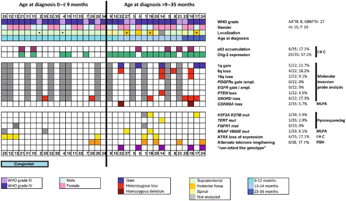
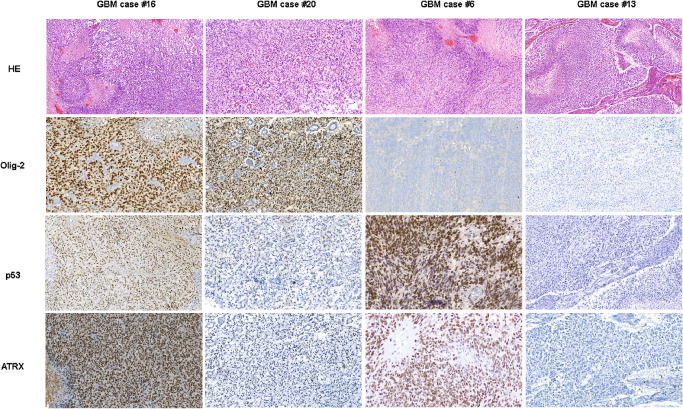
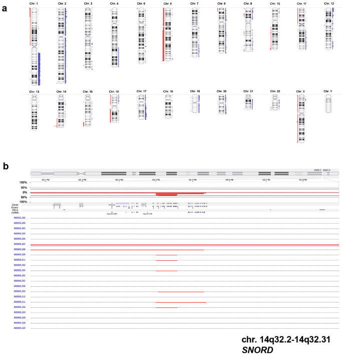
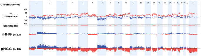
Pediatric high-grade gliomas are considered to be different when compared to adult high-grade gliomas in their pathogenesis and biological behavior. Recently, common genetic alterations, including mutations in the H3F3A/ATRX/DAXX pathway, have been described in approximately 30% of the pediatric cases. However, only few cases of infant high-grade gliomas have been analyzed so far. We investigated the molecular features of 35 infants with diffuse high-grade astrocytomas, including 8 anaplastic astrocytomas [World Health Organization (WHO) grade III] and 27 glioblastomas (WHO grade IV) by immunohistochemistry, multiplex ligation probe-dependent amplification (MLPA), pyrosequencing of glioma-associated genes and molecular inversion probe (MIP) assay. MIP and MLPA analyses showed that chromosomal alterations are significantly less frequent in infants compared with high-grade gliomas in older children and adults. We only identified H3F3A K27M in 2 of 34 cases (5.9%), with both tumors located in the posterior fossa. PDGFRA amplifications were absent, and CDKN2A loss could be observed only in two cases. Conversely, 1q gain (22.7%) and 6q loss (18.2%) were identified in a subgroup of tumors. Loss of SNORD located on chromosome 14q32 was observed in 27.3% of the infant tumors, a focal copy number change not previously described in gliomas. Our findings indicate that infant high-grade gliomas appear to represent a distinct genetic entity suggesting a different pathogenesis and biological behavior.

Keywords: ALT; ATRX; H3F3A; SNORD; glioblastoma; high-grade glioma; infants; molecular inversion probe analysis; multiplex ligation probe-dependent amplification; pediatric brain tumors.


Conflict of interest statement

The authors declare no conflict of interest.

Figures

Figure 1
Figure 1
Clinical data and molecular features of 35 infant high‐grade gliomas (iHGGs). The figure summarizes the clinical, histopathological and molecular features of the cases of iHGGs included in our study. Copy number alteration was analyzed by molecular inversion probe (MIP) analysis; genomic alteration and mutational status of candidate genes were analyzed by multiplex ligation‐dependent probe amplification (MLPA) and pyrosequencing; nuclear expression of p53 and Olig‐2 was studied by immunohistochemistry (IHC); alternative telomere lengthening (ALT) was investigated by fluorescence in situ hybridization (FISH). The patients are sorted by age (from the youngest patient, #28 on the left, to the oldest patient, #24 on the right). Median age at diagnosis was 9 months. Midline tumors are indicated by an asterisk (*). AA°III = anaplastic astrocytoma WHO grade III; ampl. = amplification; GBM°IV = glioblastoma multiforme WHO grade IV; WHO = World Health Organization.
Figure 2
Figure 2
Histopathological and immunohistochemical features (p53, Olig‐2 and ATRX) of four representative cases of infant glioblastoma patients. Glioblastomas [glioblastoma multiforme (GBM)] show microvascular proliferation and tumor necrosis with pseudopalisading of the vital tumor cells.
Figure 3
Figure 3
Virtual karyogram and loss of SNORD in 22 tumors analyzed by molecular inversion probe (MIP) arrays. Gains of chromosome 1q or losses of chromosome 6q become obvious (A). Losses are indicated in red and gains in blue. As a focal genetic alteration, a heterozygous loss of SNORD on chromosome region 14q32, a known tumor‐suppressor region, could be detected in six cases (B).
Figure 4
Figure 4
Cumulative chromosomal aberrations of 22 infant cases in comparison to 16 non‐infant pediatric high‐grade gliomas. Molecular inversion probe (MIP) analysis shows that chromosomal alterations are significantly less frequent in diffuse infant high‐grade gliomas (iHGGs) compared to pediatric high‐grade gliomas (pHGGs). The comparison plot is obtained by subtracting the genomic alterations of groups. The difference is expressed in percentage for the gains and losses for each location (gain and loss are shown as up and down, respectively) and tracked in the upper part of the figure. Bars in the track “Significant” indicate regions where there is a significant difference of the copy number changes between the compared groups (P < 0.05). The profile of copy number changes (losses in red, gains in blue) for each group is displayed in the lower track of the plot.
Figure 5
Figure 5
Box plot diagrams of the comparison of infants vs. non‐infant high‐grade gliomas (HGGs) using a Mann–Whitney rank sum test. The graphs show the number of chromosomes with cytogenetic alterations (gain/losses) affecting the whole chromosomes (A), the number of chromosome arms with cytogenetic alterations (gains/losses) affecting the entire chromosome arm (B) and number of chromosome arms with focal copy number aberrations (C) in infant high‐grade gliomas (iHGGs) compared to pediatric high‐grade gliomas (pHGGs). The iHGGs presented a significantly lower frequency of chromosomal alterations when compared to their non‐infant counterparts. The horizontal line represents the median value; the lower and upper limits of the box represent the interquartile range. Focal copy number aberrations are defined as gains or losses spanning from 10% to 50% of a chromosome arm.

References

    1. Abedalthagafi M, Phillips JJ, Kim GE, Mueller S, Haas‐Kogen DA, Marshall RE et al (2013) The alternative lengthening of telomere phenotype is significantly associated with loss of ATRX expression in high‐grade pediatric and adult astrocytomas: a multi‐institutional study of 214 astrocytomas. Mod Pathol 26:1425–1432. - PMC - PubMed
    1. Antonelli M, Buttarelli FR, Arcella A, Nobusawa S, Donofrio V, Oghaki H, Giangaspero F (2010) Prognostic significance of histological grading, p53 status, YKL‐40 expression, and IDH1 mutations in pediatric high‐grade gliomas. J Neurooncol 5:209–215. - PubMed
    1. Batra V, Sands SA, Holmes E, Russell Geyer J, Yates A, Becker L et al (2014) Long‐term survival of children less than six years of age enrolled on the CCG‐945 phase III trial for newly‐diagnosed high‐grade glioma: a report from the Children's Oncology Group. Pediatr Blood Cancer 61:151–157. - PMC - PubMed
    1. Bax DA, Mackay A, Little SE, Carvalho D, Viana‐Pereira M, Tamber N et al (2010) A distinct spectrum of copy number aberrations in pediatric high‐grade gliomas. Clin Cancer Res 16:3368–3377. - PMC - PubMed
    1. Beroukhim R, Getz G, Nghiemphu L, Barretina J, Hsueh T, Linhart D et al (2007) Assessing the significance of chromosomal aberrations in cancer: methodology and application to glioma. Proc Natl Acad Sci U S A 104:20007–20012. - PMC - PubMed

MeSH terms