. 2014 Sep 18;16(5):438.
doi: 10.1186/s13058-014-0438-2.

A novel role for ezrin in breast cancer angio/lymphangiogenesis

A novel role for ezrin in breast cancer angio/lymphangiogenesis

Abdi Ghaffari et al. Breast Cancer Res. .

Erratum in

Abstract

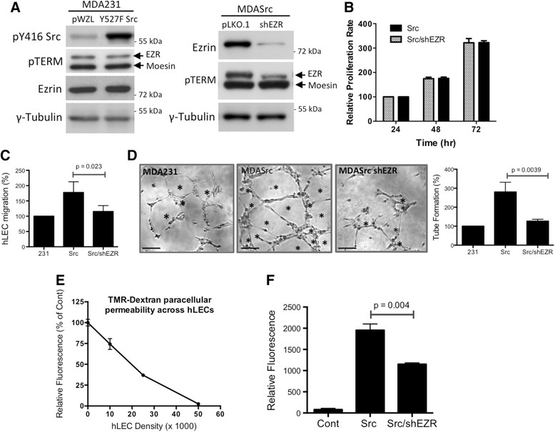
Introduction: Recent evidence suggests that tumour lymphangiogenesis promotes lymph node metastasis, a major prognostic factor for survival of breast cancer patients. However, signaling mechanisms involved in tumour-induced lymphangiogenesis remain poorly understood. The expression of ezrin, a membrane cytoskeletal crosslinker and Src substrate, correlates with poor outcome in a diversity of cancers including breast. Furthermore, ezrin is essential in experimental invasion and metastasis models of breast cancer. Ezrin acts cooperatively with Src in the regulation of the Src-induced malignant phenotype and metastasis. However, it remains unclear if ezrin plays a role in Src-induced tumour angio/lymphangiogenesis.

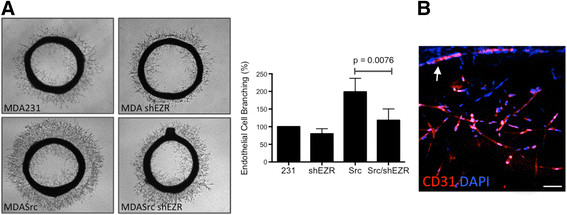
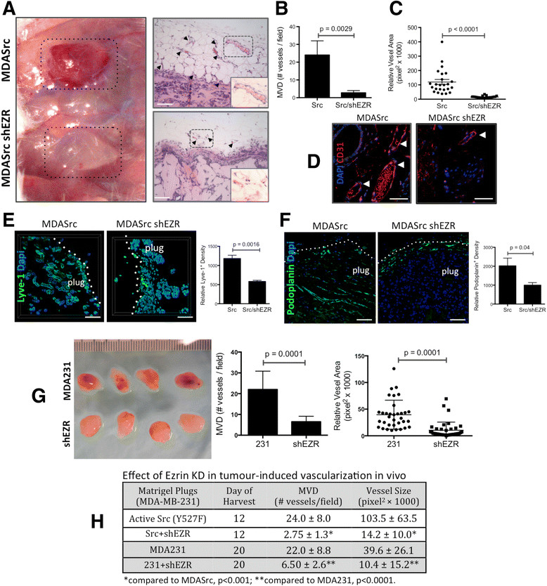
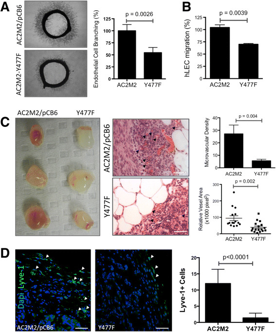
Methods: The effects of ezrin knockdown and mutation on angio/lymphangiogenic potential of human MDA-MB-231 and mouse AC2M2 mammary carcinoma cell lines were examined in the presence of constitutively active or wild-type (WT) Src. In vitro assays using primary human lymphatic endothelial cells (hLEC), an ex vivo aortic ring assay, and in vivo tumour engraftment were utilized to assess angio/lymphangiogenic activity of cancer cells.

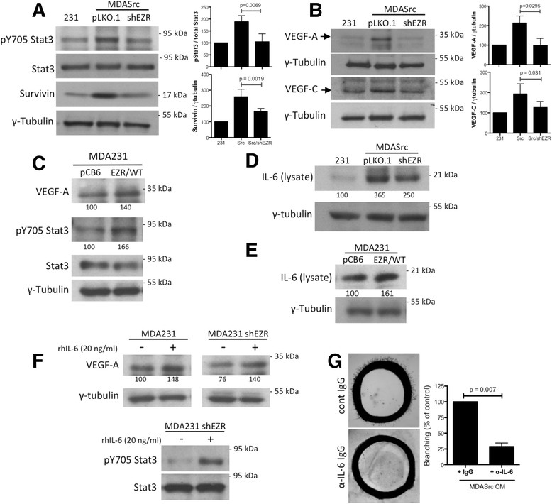
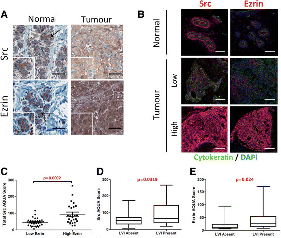
Results: Ezrin-deficient cells expressing activated Src displayed significant reduction in endothelial cell branching in the aortic ring assay in addition to reduced hLEC migration, tube formation, and permeability compared to the controls. Intravital imaging and microvessel density (MVD) analysis of tumour xenografts revealed significant reductions in tumour-induced angio/lymphangiogenesis in ezrin-deficient cells when compared to the WT or activated Src-expressing cells. Moreover, syngeneic tumours derived from ezrin-deficient or Y477F ezrin-expressing (non-phosphorylatable by Src) AC2M2 cells further confirmed the xenograft results. Immunoblotting analysis provided a link between ezrin expression and a key angio/lymphangiogenesis signaling pathway by revealing that ezrin regulates Stat3 activation, VEGF-A/-C and IL-6 expression in breast cancer cell lines. Furthermore, high expression of ezrin in human breast tumours significantly correlated with elevated Src expression and the presence of lymphovascular invasion.

Conclusions: The results describe a novel function for ezrin in the regulation of tumour-induced angio/lymphangiogenesis promoted by Src in breast cancer. The combination of Src/ezrin might prove to be a beneficial prognostic/predictive biomarker for early-stage metastatic breast cancer.


Figures

Figure 1
Figure 1
Ezrin KD inhibits lymphangiogenic activity of breast cancer cells . (A) The expression levels of active Src (pY416), pT567 ezrin/pT558 moesin (pTERM) and total ezrin in MDA231, MDASrc (Y527F Src in pWZL vector) and MDASrc shEZR (shRNA in pLKO.1 vector) cells were validated by western blot. Over 85% of endogenous ezrin was depleted in our shEZR cells. (B) The relative rates of proliferation (normalized against 24 hr) of MDA231 (231), MDASrc (Src) and MDASrc shEZR (Src/shEZR) cells were assessed by an MTT assay. (C) hLEC migration assay: percent migration of hLEC, co-cultured with MDA231 (231), MDASrc (Src), or MDASrc shEZR (Src/shEZR) cells (6 hr), is presented following normalization against the MDA231-treated group. (D) Tube formation assay: hLEC were stimulated with CM from MDA231, MDASrc, and MDASrc shEZR for 4 hr prior to manual counting of the tube-like structures (marked by asterisks). The histogram displays the percentage of tube formation in each group relative to MDA231 control cells. Scale bar = 50 μm. (E) The integrity of the hLEC monolayer was assessed by the paracellular diffusion of TMR-dextran (2000 kDa). Readings are expressed in relative fluorescence as percentage of control (collagen-coated membrane alone) (n = 3). (F) Monolayer of hLECs (5 × 10⁵ cells) was co-cultured overnight with no cells (control), MDASrc, or MDASrc shEZR (shEZR). The diffusion of TMR-dextran across the hLEC barrier was measured in 90 min and presented as relative fluorescence normalized to the control (n = 4). CM: conditioned media; hLEC: human lymphatic endothelial cells; KD: knockdown; kDa: kiloDaltons; shRNA: short hairpin RNA.
Figure 2
Figure 2
Ezrin KD inhibits tumour-induced endothelial cell sprouting. (A) Aortic rings were stimulated with CM from MDA231 (231), MDA shEZR (shEZR), MDASrc (Src), and MDASrc shEZR (Src/shEZR) for 96 hr prior to imaging. Endothelial cell sprouting was quantified using ImagePro software and presented as percentage of control (bar graph; n = 4 in triplicates). ANOVA post-test analysis revealed MDASrc as the only group with significantly higher angiogenic activity. (B) Three-dimensional reconstruction of confocal images of CD31-positive endothelial cells migrating out of aortic ring (Scale bar = 200 μm). CM: conditioned media; KD: knockdown.
Figure 3
Figure 3
Ezrin KD inhibits tumour-induced angio/lymphangiogenesis in vivo . (A) Matrigel plugs containing MDASrc (Src) or MDASrc shEZR (Src/shEZR) were digitally photographed on day 12 post injection in Rag2γ mice. Tumour-induced angiogenesis was analyzed in Matrigel plugs H&E sections for (B) MVD per selected field (manual count by trained pathologist blinded to the experiment), and (C) vessel luminal area (dots represent the mean vascular area in each selected field in pixel2). (D) Nature of blood vasculature was confirmed by CD31 immunostaining. (E and F) To assess tumour-induced lymphangiogenesis, Matrigel plugs were stained for lymphatic markers Lyve-1 and podoplanin followed by quantification of relative fluorescence density of each marker (bar graphs). (G) The Matrigel plugs derived from MDA231 (231) and MDA shEZR (shEZR) were harvested on day 20 post injection and assessed for MVD and vessel lumen area in corresponding H&E sections. (H) The effect of ezrin KD on MVD and vessel lumen size in tumours derived from MDA231 and MDASrc harvested on days 12 and 20 is summarized. Arrowheads in panels A and D point to microvessels in the tumour H&E sections. Inserts in panel A represent higher magnifications (×400) of areas in dotted boxes. P values were obtained from unpaired t test statistical analysis. All scale bars = 200 μm, except panel D bar = 50 μm. H&E: hematoxylin and eosin; KD: knockdown; MVD: microvessel density; Rag2γ: Rag2-/-II2rg-/- mice.
Figure 4
Figure 4
Expression of Y477F mutant ezrin abrogates tumour-induced angio/lymphangiogenesis. (A) Aortic rings were stimulated with CM from AC2M2 cells expressing pCB6 empty vector or Y477F ezrin and endothelial sprouting imaged (96 hr) and analyzed by ImagePro software (n = 3, bar graph). (B) Transwell migration of hLEC co-cultured with AC2M2 or Y477F cells (6 hr) was assessed by automatic count (ImageJ) of cells stained with DAPI. (C) Syngeneic Matrigel plug assay of AC2M2 or Y477F cells in CBA/J mice photographed on day 12 post injection along with representative H&E sections (arrows point to microvessels). Angiogenic activity was analyzed by MVD and vessel cross-sectional area (graphs to the right). (D) Lyve-1-positive cells (arrows) in plugs were quantified in selected hot spots by pathologist blinded to the study. P values were obtained from unpaired t test statistical analysis. Scale bars = 50 μm. CM: conditioned media; H&E: hematoxylin and eosin; MVD: microvessel density.
Figure 5
Figure 5
Ezrin KD blocks Stat3 phosphorylation, VEGF-A/-C, and IL-6 expression. Representative western blots display pY705-Stat3, Stat3, and survivin (panel A) and VEGF-A/-C (panel B) levels in MDA231, MDASrc, and MDASrc shEZR lysates. γ-tubulin expression was used as loading control. Densitometry results are presented as an average (n = 3) ratio of target protein to γ-tubulin and normalized against MDA231 controls (bar graphs). P values were calculated using unpaired t test. (C) Cell lysates from MDA231 cells expressing the empty vector pCB6 or wild-type ezrin (EZR/WT) were analyzed for VEGF-A, pY705 Stat3, and Stat3 levels. (D) Immunoblot analysis of IL-6 in cell lysates from MDA231 and MDASrc cells expressing the empty vector pLKO.1 or shEZR vector. (E) Immunoblot analysis of IL-6 levels in cell lysate from MDA231 cells expressing the empty vector pCB6 or wild-type ezrin (EZR/WT). (F) Immunoblot analysis of VEGF-A, pY705 Stat3, and Stat3 in MDA231 shEZR cells treated with vehicle alone or 20 ng/ml recombinant human IL-6 for 20 hr. (G) The angiogenic activity of CM collected from confluent MDASrc in the presence of α-IL-6 neutralizing antibody or non-immune IgG was assessed using the aortic ring assay. Endothelial cell sprouting was quantified using ImagePro software and presented as percentage of control (bar graph). Identical amount of α-IL-6 antibody was added to rings in the MDASrc group to control for the direct effect of antibody on endothelial cell sprouting. Panels C-F: relative densitometry values are displayed under corresponding bands (normalized against γ-tubulin). CM: conditioned media; IgG: immunoglobulin G; IL-6: interleukin-6; KD: knockdown; Stat3: signal transducer and activator of transcription 3; VEGF: vascular endothelial growth factor.
Figure 6
Figure 6
Src and ezrin expression in human breast cancer. (A) IHC staining of breast normal and tumour tissue was used to validate Src and ezrin antibodies. Scale bars = 75 μm. (B) Cy5 IF staining was used to assess the expression levels of Src and ezrin in the 63-patient breast cancer TMA. Representative tumour cores with high and low expression of Src and ezrin are presented. Images were acquired using Aperio ScanScopeFL. Scale bars = 100 μm. (C) Association between dichotomized ezrin AQUA scores (cutoff: median) and continuous Src AQUA scores based on IF-stained 63-TMA. (D, E) Continuous AQUA scores of total Src and ezrin were correlated against dichotomized LVI status. Statistical associations were assessed using the unpaired Student's t test. Associations were considered significant when P <0.05. AQUA: automated quantification analysis IF: immunofluorescence; IHC: immunohistochemistry; LVI: lymphovascular involvement; TMA: tissue microassay.

Similar articles

Cited by

References

    1. Fehon RG, McClatchey AI, Bretscher A. Organizing the cell cortex: the role of ERM proteins. Nat Rev Mol Cell Biol. 2010;11:276–287. doi: 10.1038/nrm2866. - DOI - PMC - PubMed
    1. Li L, Wang YY, Zhao ZS, Ma J. Ezrin is associated with gastric cancer progression and prognosis. Pathol Oncol Res. 2011;17:909–915. doi: 10.1007/s12253-011-9402-y. - DOI - PubMed
    1. Sarrio D, Rodriguez-Pinilla SM, Dotor A, Calero F, Hardisson D, Palacios J. Abnormal ezrin localization is associated with clinicopathological features in invasive breast carcinomas. Breast Cancer Res Treat. 2006;98:71–79. doi: 10.1007/s10549-005-9133-4. - DOI - PubMed
    1. Arslan AA, Silvera D, Arju R, Giashuddin S, Belitskaya-Levy I, Formenti SC, Schneider RJ. Atypical ezrin localization as a marker of locally advanced breast cancer. Breast Cancer Res Treat. 2012;134:981–988. doi: 10.1007/s10549-012-2017-5. - DOI - PubMed
    1. Bruce B, Khanna G, Ren L, Landberg G, Jirstrom K, Powell C, Borczuk A, Keller ET, Wojno KJ, Meltzer P, Baird K, McClatchey A, Bretscher A, Hewitt SM, Khanna C. Expression of the cytoskeleton linker protein ezrin in human cancers. Clin Exp Metastasis. 2007;24:69–78. doi: 10.1007/s10585-006-9050-x. - DOI - PubMed

Publication types

MeSH terms

LinkOut - more resources