. 2014 Nov 7;289(45):31224-40.
doi: 10.1074/jbc.M114.559237. Epub 2014 Sep 17.

Interplay between sumoylation and phosphorylation for protection against α-synuclein inclusions

Affiliations

Interplay between sumoylation and phosphorylation for protection against α-synuclein inclusions

Hedieh Shahpasandzadeh et al. J Biol Chem. .

Abstract

Parkinson disease is associated with the progressive loss of dopaminergic neurons from the substantia nigra. The pathological hallmark of the disease is the accumulation of intracytoplasmic inclusions known as Lewy bodies that consist mainly of post-translationally modified forms of α-synuclein. Whereas phosphorylation is one of the major modifications of α-synuclein in Lewy bodies, sumoylation has recently been described. The interplay between α-synuclein phosphorylation and sumoylation is poorly understood. Here, we examined the interplay between these modifications as well as their impact on cell growth and inclusion formation in yeast. We found that α-synuclein is sumoylated in vivo at the same sites in yeast as in human cells. Impaired sumoylation resulted in reduced yeast growth combined with an increased number of cells with inclusions, suggesting that this modification plays a protective role. In addition, inhibition of sumoylation prevented autophagy-mediated aggregate clearance. A defect in α-synuclein sumoylation could be suppressed by serine 129 phosphorylation by the human G protein-coupled receptor kinase 5 (GRK5) in yeast. Phosphorylation reduced foci formation, alleviated yeast growth inhibition, and partially rescued autophagic α-synuclein degradation along with the promotion of proteasomal degradation, resulting in aggregate clearance in the absence of a small ubiquitin-like modifier. These findings suggest a complex interplay between sumoylation and phosphorylation in α-synuclein aggregate clearance, which may open new horizons for the development of therapeutic strategies for Parkinson disease.

Keywords: Autophagy; Post-translational Modification; Proteasome; Yeast; α-Synuclein.


Figures

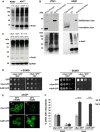
FIGURE 1.
FIGURE 1.
αSyn is sumoylated in S. cerevisiae and impairment of sumoylation increases αSyn toxicity and foci formation. A, total protein extract of ulp1 temperature-sensitive yeast cells, defective in SUMO deconjugation, co-expressing integrated αSyn (driven by GAL1 promoter at TRP1 locus) and the His6-tagged yeast SUMO protein Smt3 (driven by ADH1 promoter at HIS3 locus). Enriched sumoylated proteins in the ulp1ts strain in comparison with the control W303 were detected by Western blot with Smt3 antibody (α-Smt3). EV, yeast cells, transformed with empty vector. B, nickel pulldown of His6-tagged yeast SUMO protein Smt3 (His6-smt3) in ulp1ts cells co-expressing αSyn. Sumoylated αSyn (αSyn and A30PSyn) was detected in the pulldown fractions with αSyn antibody (upper panel). Unmodified αSyn was detected in the flow-through. Yeast cells transformed with empty vector were used as a control. Western hybridization of the same blot with Smt3 antibody (lower panel) verified the Ni2+ pulldown (lower panel). C, Western hybridization with Smt3 antibody of total protein extract of smt3 temperature-sensitive yeast cells, co-expressing αSyn or empty vector (EV) at permissive (25 °C) or restrictive temperature (30 °C). D, spotting assay of conditional smt3ts mutant strain expressing αSyn-GFP or A30P-GFP at permissive (25 °C; +SUMO; Smt3 functional) or restrictive temperature (30 °C; −SUMO; Smt3 dysfunctional). GAL1-driven αSyn-GFP is expressed from two genomically integrated copies. GAL1-driven A30P-GFP is expressed from a 2-μm plasmid. GFP, expressed from the same promoter, is used as a control. Yeast cells were spotted in 10-fold dilutions on selection plates containing glucose (αSyn 'OFF') or galactose (αSyn 'ON'). E, fluorescence microscopy of smt3ts cells expressing αSyn-GFP or A30P-GFP at permissive (25 °C; +SUMO) or restrictive temperature (30 °C; −SUMO). Scale bar, 1 μm. F, quantification of the percentage of cells displaying αSyn inclusions. W303 cells expressing αSyn-GFP or A30P-GFP at 25 or 30 °C were used in comparison with smt3ts cells expressing αSyn-GFP or A30P-GFP at permissive (25 °C; +SUMO) or restrictive temperature (30 °C; −SUMO). At least 300 cells were counted per strain and experiment. Significance of differences was calculated with t test (***, p < 0.001, n = 3).
FIGURE 2.
FIGURE 2.
Lysine 96 and 102 are conserved as major sumoylation sites of αSyn in eukaryotes. A, lysine to arginine substitutions at positions 96 and 102 resulted in decreased αSyn sumoylation. αSyn and A30PSyn and the corresponding αSyn amino acid variants K96R/K102R were transformed into ulp1ts yeast cells expressing the yeast SUMO protein His6-Smt3. His6-tagged SUMO conjugates were pulled down by Ni2+-NTA. αSyn was detected by Western hybridization using αSyn antibody (upper panel). Western hybridization of the same blot with Smt3 antibody (α-Smt3) verified the Ni2+-NTA pulldown (lower panel). B, spotting assay of W303 yeast cells, carrying two copies of GAL1-driven αSyn-GFP and K96R/K102R-GFP. Yeast cells were spotted in 10-fold dilutions on selection plates containing glucose (αSyn 'OFF') or galactose (αSyn 'ON'). C, quantification of the percentage of cells displaying αSyn inclusions in W303 yeast background. Significance of differences was calculated with t test (***, p < 0.001, n = 3).
FIGURE 3.
FIGURE 3.
Expression of the human kinases GRK5/PLK2 increases αSyn Ser-129 phosphorylation in yeast. A, Smt3ts mutant cells co-expressing αSyn and GRK5 or PLK2 at permissive (25 °C; +SUMO) or restrictive temperature (30 °C; −SUMO). The phosphorylation level of αSyn on Ser-129 was detected by αSyn Ser-129 phosphorylation-specific antibody (αSyn pSer129) when expressed either alone (αSyn-GFP + empty vector (EV)) or in the presence of GRK5 or PLK2. Immunoblotting analysis of yeast cells expressing S129A-GFP with αSyn Ser(P)129 antibody (right panel) was used as a control for antibody specificity. B, quantification of αSyn Ser-129 phosphorylation level in the presence or absence of GRK5 and PLK2, respectively, at permissive (25 °C; +SUMO) or restrictive temperature (30 °C; −SUMO). Densitometric analysis of the immunodetection of αSyn Ser(P)-129 was normalized to the total amount of αSyn and relative to αSyn + EV at permissive temperature (25 °C; +SUMO). Significance of differences was calculated with one-way ANOVA with Bonferroni's multiple comparison test (**, p < 0.01; ##, p < 0,05 versus empty vector, n = 4). C, Western hybridization of W303 yeast cells co-expressing K96R/K102R-GFP and GRK5 or PLK2. The phosphorylation level of sumoylation-deficient αSyn mutant on Ser-129 was visualized with αSyn Ser(P)-129. D, quantification of αSyn Ser-129 phosphorylation levels of sumoylation-deficient αSyn mutant in the presence or absence of GRK5 and PLK2. Densitometric analysis of the immunodetection of αSyn Ser(P)-129 was normalized to the total amount of αSyn. Significance of differences was calculated with one-way ANOVA (**, p < 0.01, n = 4).
FIGURE 4.
FIGURE 4.
Increased αSyn Ser-129 phosphorylation level by GRK5/PLK2 expression alleviates the toxicity and reduces foci formation associated with impaired sumoylation in smt3ts cells. A, spotting assay of smt3ts cells co-expressing αSyn-GFP or S129A-GFP with GRK5 or PLK2 either at permissive (25 °C; +SUMO) or restrictive temperature (30 °C; −SUMO). Yeast cells were spotted in 10-fold dilutions on selection plates containing glucose (αSyn 'OFF'; kinases 'OFF') or galactose (αSyn 'ON'; kinases 'ON'). B, spotting assay of W303 yeast cells, carrying two copies of GAL1-driven αSyn-GFP and K96R/K102R-GFP in the presence of GRK5 and PLK2 or empty vector. Yeast cells were spotted in 10-fold dilutions on selection plates containing glucose (αSyn 'OFF'; kinases 'OFF') or galactose (αSyn 'ON'; kinases 'ON'). C, fluorescence microscopy of smt3ts cells expressing αSyn in the presence or absence of GRK5 or PLK2 (left panel). Scale bar, 1 μm. Quantification of percentage of cells displaying αSyn inclusions in the presence or absence of GRK5/PLK2 (upright) and quantification of cells with foci formation expressing S129A-GFP with and without overexpression of GRK5/PLK2. Significance of differences was calculated with t test with respect to αSyn-GFP at the same temperature (**, p < 0.01, n = 3).
FIGURE 5.
FIGURE 5.
αSyn aggregate clearance upon promoter shutoff. A, inhibition of autophagy by deletion of ATG1 and ATG7. Expression of αSyn-GFP and K96R/K102R-GFP was induced for 4 h in galactose medium and then the cells were shifted to glucose medium. Quantification of the reduction of inclusions was done 2 h after the promoter shutoff and was presented as the ratio of aggregate clearance in the deletion strain to aggregate clearance in the isogenic wild-type strain. Significance of differences was calculated with t test (**, p < 0.01, n = 3). B, inhibition of the vacuolar degradation pathway by PMSF. Quantification was of cells expressing αSyn-GFP, K96R/K102R-GFP, and K96R/K102R-GFP and co-expressing GRK5 or PLK2, respectively. αSyn-GFP and K96R/K102R-GFP were expressed from two genomically integrated copies. After 4 h of induction of the protein expression in galactose medium, cells were shifted to glucose medium supplemented with 1 mm PMSF dissolved in ethanol (EtOH) or only EtOH as a control. Quantification of the reduction of inclusions was done 2 h after the promoter shutoff. Cells with inclusions were counted and presented as a ratio to the control (EtOH). C, inhibition of the proteasome with MG132. The protein expression was induced as above, and the cells were shifted to glucose medium supplemented with 75 μm MG132 and dissolved in DMSO or only DMSO as a control. Quantification of the reduction of inclusions was done 2 h after the promoter shutoff. Cells with inclusions were counted and presented as a ratio to the control (DMSO). Significance of differences was calculated with one-way ANOVA with Bonferroni's multiple comparison test (**, p < 0.01; ***, p < 0.001; n = 3; #, p < 0.05 versus K96R/K102R-GFP) (Bonferroni's multiple comparison test).
FIGURE 6.
FIGURE 6.
αSyn aggregate clearance in ulp1ts cells. A, percentage of cells displaying αSyn-GFP inclusions after 6 h of induction of ulp1ts cells, expressing αSyn-GFP or K96R/K102R-GFP. B, spotting assay of ulp1ts cells expressing αSyn-GFP or K96R/K102R-GFP from 2 μm plasmid. Yeast cells were spotted in 10-fold dilutions on selection plates containing glucose (αSyn 'OFF') or galactose (αSyn 'ON'). C, fluorescence microscopy of ulp1ts cells expressing αSyn-GFP or K96R/K102R-GFP. After 6 h of induction of the protein expression in galactose medium, cells were shifted to glucose medium. Scale bar, 1 μm. D, quantification of the reduction of inclusions 2 h after the promoter shutoff. Cells with inclusions were counted and presented as a ratio to time point 0 h. Significance of differences was calculated with t test (**, p < 0.01, n = 3).
FIGURE 7.
FIGURE 7.
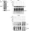
αSyn is ubiquitinated in yeast cells. A, αSyn-His6 protein was purified by Ni2+ pulldown and subjected to immunoprecipitation with ubiquitin antibody. The ubiquitinated and phosphorylated αSyn was detected by αSyn and αSyn Ser(P)-129-specific antibody, respectively. Empty vector (EV) was used as a control. B, Smt3ts cells expressing αSyn-His6 co-transformed with GRK5 or PLK2 and empty vector of the kinases (EV) as a control at permissive (25 °C; +SUMO) or restrictive temperature (30 °C; −SUMO). The purified αSyn protein from Ni2+ pulldown was subjected to ubiquitin immunoprecipitation (IP Ubi). As a control, the same experiments were performed without addition of ubiquitin antibody. The ubiquitinated αSyn was analyzed by Western hybridization with an antibody against αSyn. Western hybridization of the same blots after stripping with ubiquitin antibody (lower panels). A representative result is shown from three independent experiments.
FIGURE 8.
FIGURE 8.
Effect of sumoylation and increased αSyn Ser-129 phosphorylation by GRK5/PLK2 on αSyn protein stability. GAL1 promoter shutoff studies and drug treatments. A, Smt3ts yeast cells expressing αSyn with or without GRK5 or PLK2 at permissive temperature (25 °C) were induced for 4 h in galactose (αSyn on) and then transferred to glucose containing medium (αSyn off). Immunoblotting analysis was performed at the indicated time points after promoter shutoff with αSyn antibody and GAPDH antibody as loading control. B, W303 cells expressing αSyn, K96R/K102R (C), K96R/K102R + GRK5 (D), or K96R/K102R + PLK2 (E) were induced for 4 h in galactose (αSyn on) and then transferred to glucose containing medium (αSyn off). The glucose medium was supplemented with 75 μm MG132 or 1 mm PMSF. Immunoblotting analysis was performed at the indicated time points after promoter shutoff with αSyn antibody and GAPDH antibody as loading control. A representative result is shown from three independent experiments. Right panels, densitometric analysis of the immunodetection of αSyn-GFP relative to the GAPDH loading control. Significance of differences was calculated with one-way ANOVA with Bonferroni's multiple comparison test (***, p < 0.001; **, p < 0.01; *, p < 0.05; ##, p < 0.05 versus 0 h (Bonferroni's multiple comparison test)).
FIGURE 9.
FIGURE 9.
Inhibition of the proteasome with MG132. Promoter shutoff of yeast cells, expressing αSyn-GFP in the absence and presence of proteasome inhibitor MG132. Representative Western hybridization with ubiquitin antibody, showing accumulation of ubiquitinated species after treatment with MG132 in comparison with the control (no drug and 0 h of treatment).
FIGURE 10.
FIGURE 10.
Effect of sumoylation and GRK5/PLK2 expression on S129A-GFP protein stability. GAL1 promoter shutoff studies. A, Smt3ts yeast cells expressing S129A-GFP with or without GRK5 or PLK2 at permissive temperature (25 °C, +SUMO) and restrictive temperature (30 °C, −SUMO) were induced for 4 h in galactose (αSyn on) and then transferred to glucose-containing medium (αSyn off). Immunoblotting analysis was performed at the indicated time points after promoter shutoff with αSyn antibody and GAPDH antibody as loading control. A representative result is shown from three independent experiments. B, densitometric analysis of the immunodetection of S129A-GFP relative to the GAPDH loading control. Significance of differences was calculated with one-way ANOVA with Bonferroni's multiple comparison test (**, p < 0.01; ##, p < 0.05 versus 0 h (Bonferroni's multiple comparison test).
FIGURE 11.
FIGURE 11.
αSyn clearance and degradation in yeast. Proteasome and autophagy/vacuole as major degradation pathways are depicted. When synthesis of αSyn is switched off, wild-type yeast cells clear αSyn aggregates within hours and regain normal growth rates (38). In the presence of functional SUMO, the aggregates are primarily cleared by the autophagy/vacuolar pathway. When sumoylation is impaired, the aggregate clearance through autophagy/vacuolar pathway is prevented, and the proteasomal degradation is promoted. Increase of Ser-129 phosphorylation level by GRK5 or PLK2 rescues the autophagic aggregate clearance and additionally promotes the proteasomal degradation. For monomers, degradation of soluble αSyn monomers occurs through both pathways. Inhibition of αSyn sumoylation has a strong effect on monomer protein stability, significantly increasing the half-life of the protein and inhibiting the degradation through both pathways. Phosphorylation at Ser-129 by GRK5 or PLK2 decreases the protein stability and promotes degradation of soluble αSyn through proteasome and autophagy pathways.

Similar articles

Cited by

References

    1. Obeso J. A., Rodriguez-Oroz M. C., Goetz C. G., Marin C., Kordower J. H., Rodriguez M., Hirsch E. C., Farrer M., Schapira A. H., Halliday G. (2010) Missing pieces in the Parkinson's disease puzzle. Nat. Med. 16, 653–661 - PubMed
    1. Galvin J. E., Lee V. M., Trojanowski J. Q. (2001) Synucleinopathies: clinical and pathological implications. Arch. Neurol. 58, 186–190 - PubMed
    1. Forno L. S., DeLanney L. E., Irwin I., Langston J. W. (1996) Electron microscopy of Lewy bodies in the amygdala-parahippocampal region. Comparison with inclusion bodies in the MPTP-treated squirrel monkey. Adv. Neurol. 69, 217–228 - PubMed
    1. Spillantini M. G., Schmidt M. L., Lee V. M., Trojanowski J. Q., Jakes R., Goedert M. (1997) α-Synuclein in Lewy bodies. Nature 388, 839–840 - PubMed
    1. Polymeropoulos M. H., Lavedan C., Leroy E., Ide S. E., Dehejia A., Dutra A., Pike B., Root H., Rubenstein J., Boyer R., Stenroos E. S., Chandrasekharappa S., Athanassiadou A., Papapetropoulos T., Johnson W. G., Lazzarini A. M., Duvoisin R. C., Di Iorio G., Golbe L. I., Nussbaum R. L. (1997) Mutation in the α-synuclein gene identified in families with Parkinson's disease. Science 276, 2045–2047 - PubMed

Publication types

LinkOut - more resources